. 2015 Apr 9:2:36-43.
doi: 10.1016/j.bonr.2015.03.003. eCollection 2015 Jun.

Mineral trioxide aggregate induces osteoblastogenesis via Atf6

Affiliations

Mineral trioxide aggregate induces osteoblastogenesis via Atf6

Toyonobu Maeda et al. Bone Rep. .

Abstract

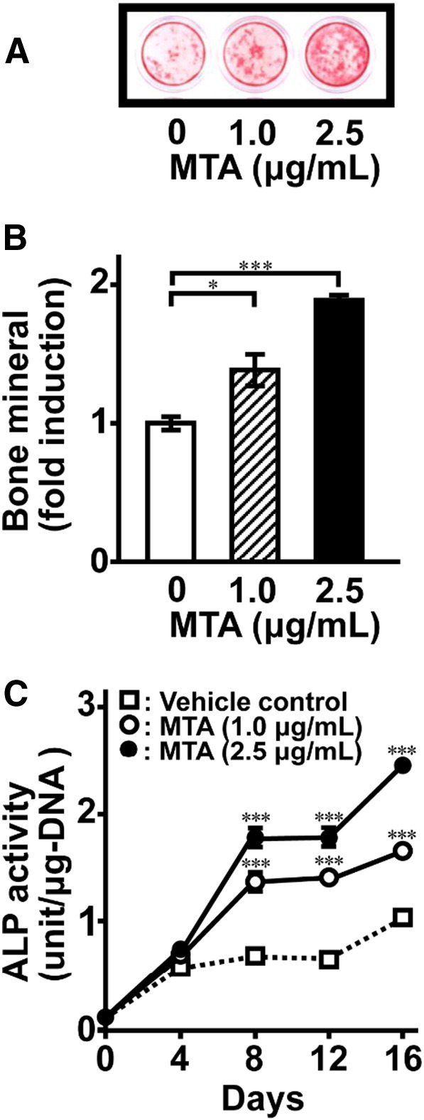
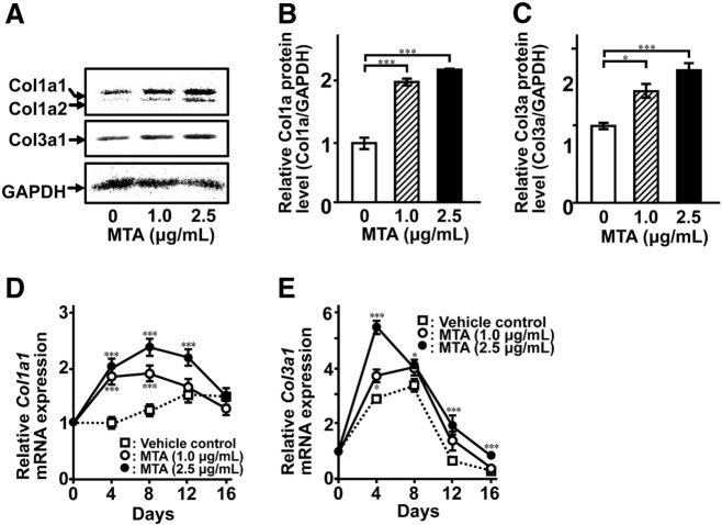
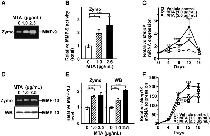
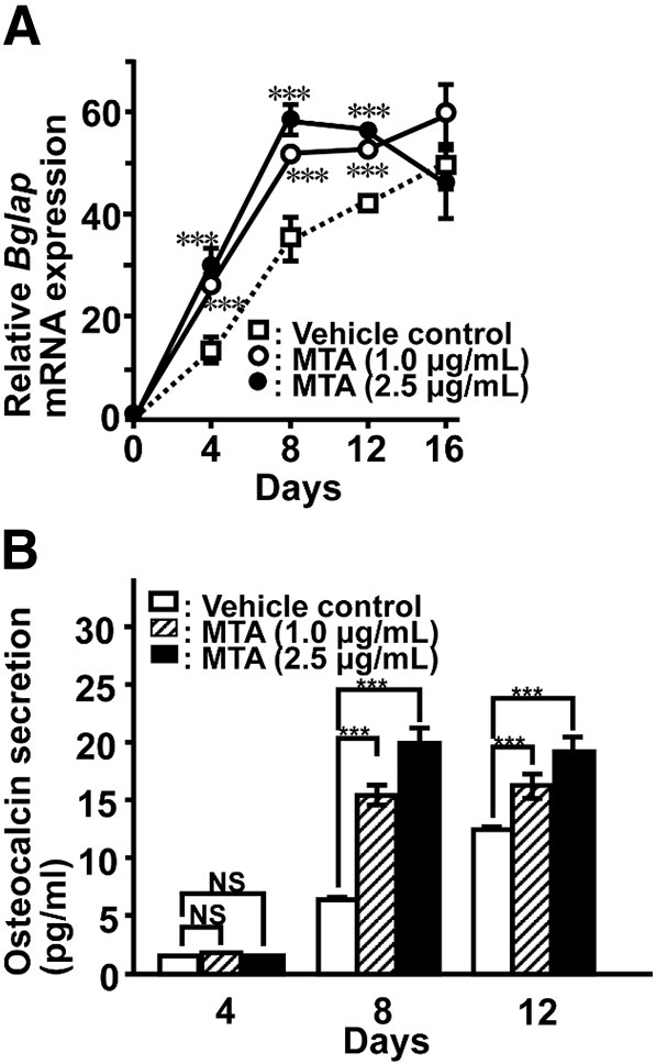
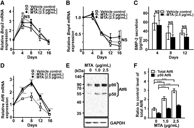
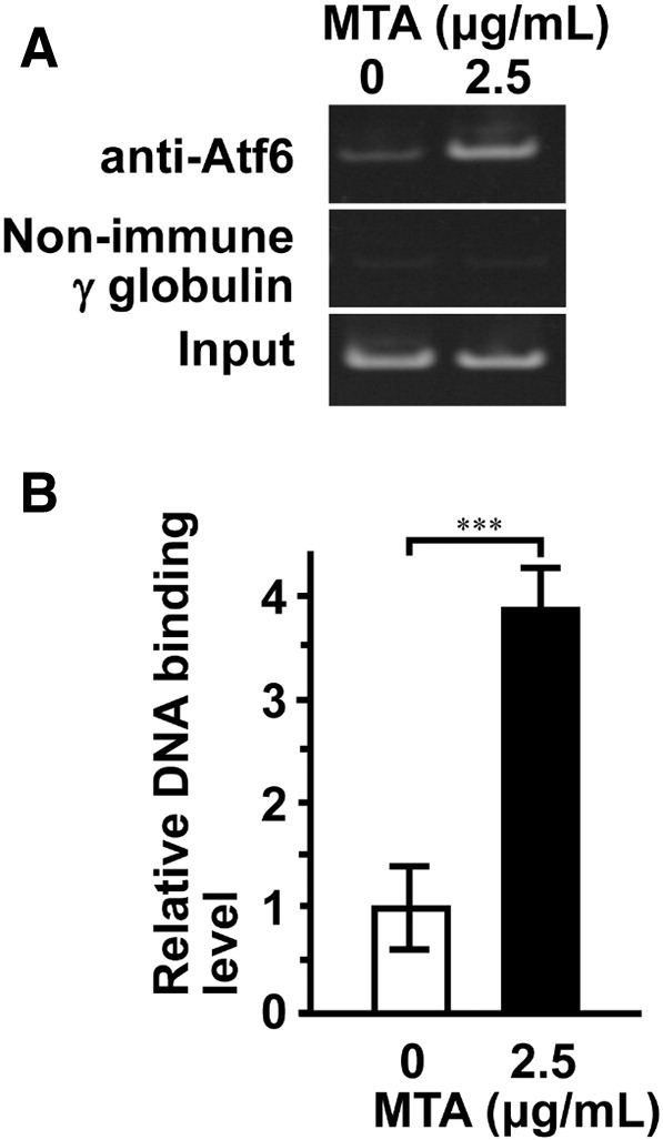
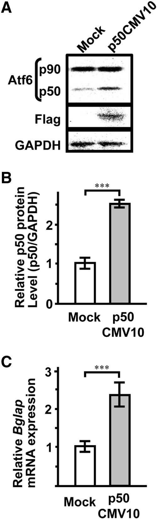
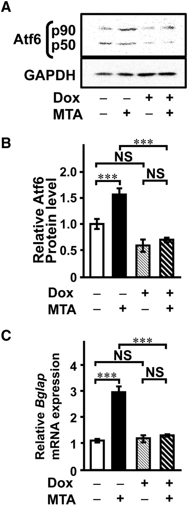
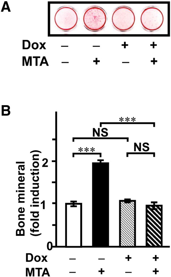
Mineral trioxide aggregate (MTA) has been recommended for various uses in endodontics. To understand the effects of MTA on alveolar bone, we examined whether MTA induces osteoblastic differentiation using MC3T3-E1 cells. MTA enhanced mineralization concomitant with alkaline phosphatase activity in a dose- and time-dependent manner. MTA increased production of collagens (Type I and Type III) and matrix metalloproteinases (MMP-9 and MMP-13), suggesting that MTA affects bone matrix remodeling. MTA also induced Bglap (osteocalcin) but not Bmp2 (bone morphogenetic protein-2) mRNA expression. We observed induction of Atf6 (activating transcription factor 6, an endoplasmic reticulum (ER) stress response transcription factor) mRNA expression and activation of Atf6 by MTA treatment. Forced expression of p50Atf6 (active form of Atf6) markedly enhanced Bglap mRNA expression. Chromatin immunoprecipitation assay was performed to investigate the increase in p50Atf6 binding to the Bglap promoter region by MTA treatment. Furthermore, knockdown of Atf6 gene expression by introduction of Tet-on Atf6 shRNA expression vector abrogated MTA-induced mineralization. These results suggest that MTA induces in vitro osteoblastogenesis through the Atf6-osteocalcin axis as ER stress signaling. Therefore, MTA in endodontic treatment may affect alveolar bone healing in the resorbed region caused by pulpal infection.

Keywords: Atf6; Bone regeneration; Endodontics; Mineral trioxide aggregate; Osteoblastgenesis.


Figures

Fig. 1
Fig. 1
MTA induces mineralization of extracellular matrix in MC3T3-E1 cells. The cells were cultured for 16 days in the absence or presence of MTA at indicated concentrations. (A) AR-S staining was performed to examine mineralized nodule formation. (B) A quantitative analysis of the intensity for AR-S staining was determined by Molecular Imager FX (Bio-Rad). (C) The time course of stimulation of ALP activity by MTA in MC3T3-E1 cells. ALP samples from whole-cell extracts were assayed using an ALP kit. Data represent the means ± SEM (n = 4). Statistical significance of differences between MTA treated group and corresponding time-matched vehicle control is indicated by *P < 0.05 and ***P < 0.001.
Fig. 2
Fig. 2
MTA induces collagen synthesis in MC3T3-E1 cells. (A) The cells were cultured for 8 days in the absence or presence of MTA at indicated concentrations. Whole-cell lysates were subjected to western blotting (A). A quantitative analysis of intensity for each immunoreacted band for Type I collagen α1 (Co1a) (B) and Type III collagen α1 (Col3a) (C) was performed by Molecular Imager FX (Bio-Rad). Total RNA was extracted, reverse-transcribed and amplified by real-time PCR with primer sets for Col1a1 and Col3a1. Data are expressed as mean ± SEM (n = 4). Statistical significance of differences between MTA treated group and corresponding time-matched vehicle control is indicated by *P < 0.05 and ***P < 0.001.
Fig. 3
Fig. 3
Production of MMPs in MTA-treated MC3T3-E1 cells. The cells were cultured in the absence or presence of MTA for 12 days. The medium was changed to serum-free conditioned medium. After 24 h, the conditioned medium was collected and concentrated. (A) MMP-9 was detected by gelatin-zymography as the precursor. (D) MMP-13 was detected by casein-zymography and western blotting as the precursor. A quantitative analysis of the intensity of each band was performed by Molecular Imager FX (Bio-Rad) (B and E). Total RNA was extracted and determined mRNA expression of Mmp9 (C) and Mmp13 (F) by real-time RT-PCR. Data represent the means ± SEM (n = 4). Statistical significance of differences between MTA treated group and corresponding time-matched vehicle control is indicated by *P < 0.05, **P < 0.01 and ***P < 0.001. Zymo, zymography; WB, western blotting.
Fig. 4
Fig. 4
Time course of Bglap mRNA expression (A) and osteocalcin secretion (B) in MTA-treated MC3T3-E1. At the indicated time points, total RNA was extracted for real-time RT-PCR. To determine osteocalcin secretion, culture medium was replaced with fresh medium and the cells were further cultured with or without MTA at the indicated concentration for 24 h. Osteocalcin concentration was measured using a mouse osteocalcin ELISA kit. Data represent the means ± SEM. Statistical significance of differences between MTA treated group and corresponding time-matched vehicle control is indicated by ***P < 0.001. NS, not significant.
Fig. 5
Fig. 5
MTA induced Atf6 expression but did not BMP-2 and 4 in MC3T3-E1 cells. Total RNA was extracted, reverse-transcribed, and amplified by real-time PCR using specific primer sets for Bmp2 (A), Bmp4 (B), and Atf6 (D). (C) To determine BMP-2 protein secretion, culture medium was replaced with fresh medium at the indicated time points and the culture was continued with or without MTA for 24 h. The CM was analyzed for BMP-2 concentration using a BMP-2 ELISA Kit. (E) The cells were cultured for 12 days in the absence or presence of MTA at indicated concentration. Whole-cell lysates were subjected to immunoblotting with anti-Atf6 and GAPDH antibodies. (F) A quantitative analysis of the intensity of each band was performed by Molecular Imager FX (Bio-Rad). Total Atf6 was calculated as sum of intensities of p90 and p50 bands. Data represent the means ± SEM (n = 3). Statistical significance of differences between MTA treated group and corresponding time-matched vehicle control is indicated by ***P < 0.001. NS, not significant.
Fig. 6
Fig. 6
MTA stimulates Atf6 binding to promoter region of Bglap gene. The cells were stimulated by MTA for 4 days. ChIP assays were performed. Cell lysates were incubated with non-immune IgG control or ATF6 antibodies. (A) PCR amplification of Bglap gene promoters was performed using immunoprecipitated and non-immunoprecipitated (input) DNA by using PCR thermal cycler. (B) A quantitative study of immunoprecipitated DNA was performed by using real-time PCR. Data represent the means ± SEM (n = 3). Statistical significance of differences between MTA treated group and corresponding time-matched vehicle control is indicated by ***P < 0.001.
Fig. 7
Fig. 7
Forced expression of active Atf6 (p50Atf6) induces Bglap gene expression. The cells were transiently transfected with a p3 × FLAGp50CMV10 or mock (p3 × FLAGCMV10) in MC3T3-E1 cells and cultured for 2 days in osteogenic medium. (A) Protein level of Atf6 was analyzed by western blotting. (B) A quantitative study of intensity was performed by Molecular Imager FX (Bio-Rad). (C) Bglap (osteocalcin) mRNA expression was determined by real-time RT-PCR. Data represent the means ± SEM (n = 3). ***, P < 0.001.
Fig. 8
Fig. 8
Knockdown of Atf6 gene expression abolishes Bglap mRNA expression and mineralization by MTA. Stable clones of Atf6 shRNA were incubated with (+) or without (−) Dox and/or MTA for 16 days: MTA, 2.5 μg/mL; Dox, 1.0 μg/mL. (A) Whole-cell lysates were subjected to immunoblotting with anti-Atf6 and GAPDH antibodies on day 12 in culture. (B) A quantitative study for the intensity was performed by Molecular Imager FX (Bio-Rad). (C) Bglap mRNA expression was decreased by Atf6 shRNA. On day 12 in culture, total RNA was extracted from the cells, reverse-transcribed, and subjected to qPCR analysis. Data represent the means ± SEM (n = 3). ***, P < 0.001; NS, not significant.
Fig. 9
Fig. 9
Attenuation of Atf6 expression does not induce mineralized nodule formation in MTA-treated cells. The cells were cultured with (+) or without (−) Dox and/or MTA for 16 days: MTA, 2.5 μg/mL; Dox, 1.0 μg/mL. (A) AR-S staining was performed to examine mineralized nodule formation. (B) A quantitative study of the intensity of the AR-S staining was determined by Molecular Imager FX (Bio-Rad) (B, E). Data represent the means ± SEM (n = 3). ***, P < 0.001; NS, not significant.

References

    1. Agata Y., Katakai T., Ye S.K., Sugai M., Gonda H., Honjo T. Histone acetylation determines the developmentally regulated accessibility for T cell receptor γ gene recombination. J. Exp. Med. 2001;193:873–880. - PMC - PubMed
    1. Barthelemi S., Robinet J., Garnotel R., Antonicelli F., Schittly E., Hornebeck W. Mechanical forces-induced human osteoblasts differentiation involves MMP-2/MMP-13/MT1-MMP proteolytic cascade. J. Cell. Biochem. 2012;113:760–772. - PubMed
    1. Bernabé P.F., Azuma M.M., Ferreira L.L., Dezan-Júnior E., Gomes-Filho J.E., Cintra L.T. Root reconstructed with mineral trioxide aggregate and guided tissue regeneration in apical surgery: a 5-year follow-up. Braz. Dent. J. 2013;24:428–432. - PubMed
    1. Camilleri J. The chemical composition of mineral trioxide aggregate. J. Conserv. Dent. 2008;11:141–143. - PMC - PubMed
    1. Chen X., Shen J., Prywes R. The luminal domain of ATF6 senses endoplasmic reticulum (ER) stress and causes translocation of ATF6 from the ER to the Golgi. J. Biol. Chem. 2002;277:13045–13052. - PubMed

LinkOut - more resources