. 2015 Aug 18:3:48-56.
doi: 10.1016/j.bonr.2015.08.002. eCollection 2015 Dec.

Primary human osteoblasts with reduced alkaline phosphatase and matrix mineralization baseline capacity are responsive to extremely low frequency pulsed electromagnetic field exposure - Clinical implication possible

Affiliations

Primary human osteoblasts with reduced alkaline phosphatase and matrix mineralization baseline capacity are responsive to extremely low frequency pulsed electromagnetic field exposure - Clinical implication possible

Sabrina Ehnert et al. Bone Rep. .

Abstract

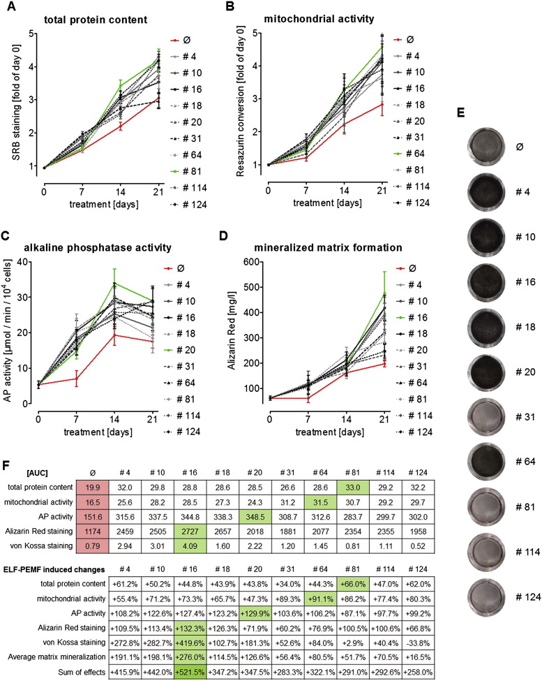
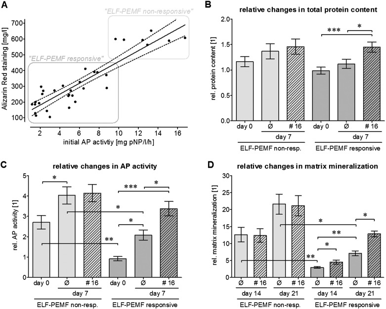
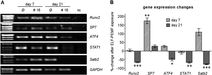
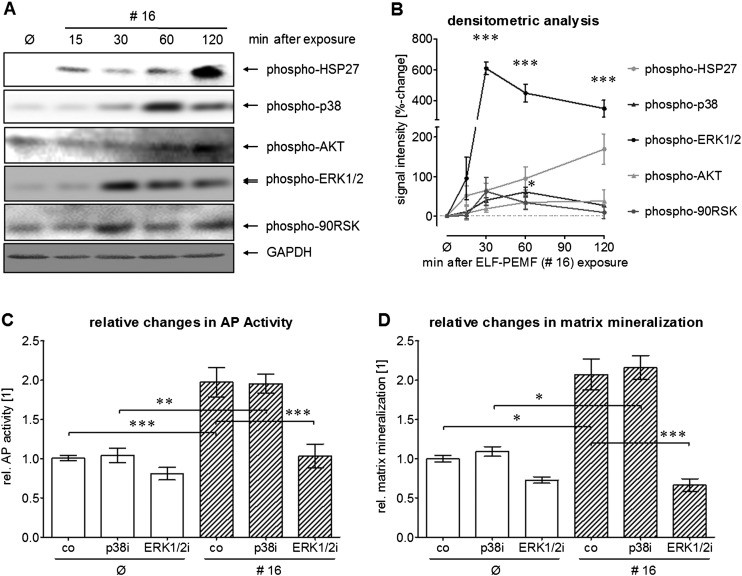
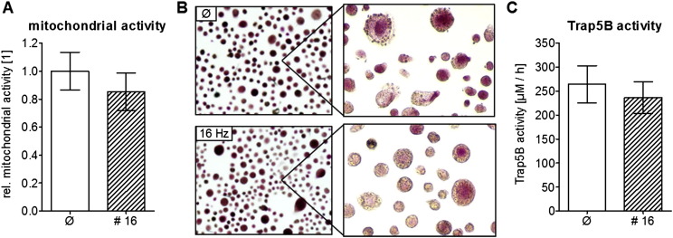
For many years electromagnetic fields (EMFs) have been used clinically with various settings as an exogenous stimulation method to promote fracture healing. However, underlying mechanisms of action and EMF parameters responsible for certain effects remain unclear. Our aim was to investigate the influence of defined EMFs on human osteoblasts' and osteoclasts' viability and function. Primary human osteoblasts and osteoclasts were treated 3 times weekly for 21 days during their maturation process using the Somagen® device (Sachtleben GmbH, Hamburg, Germany), generating defined extremely low-frequency pulsed electromagnetic fields (ELF-PEMFs). Certain ELF-PEMF treatment significantly increased the total protein content (up to 66%), mitochondrial activity (up to 91.1%) and alkaline phosphatase (AP) activity (up to 129.9%) of human osteoblasts during the entire differentiation process. Furthermore, ELF-PEMF treatment enhanced formation of mineralized matrix (up to 276%). Interestingly, ELF-PEMF dependent induction of AP activity and matrix mineralization was strongly donor dependent - only osteoblasts with a poor initial osteoblast function responded to the ELF-PEMF treatment. As a possible regulatory mechanism, activation of the ERK1/2 signaling pathway was identified. Maturation of osteoclasts from human monocytes was not affected by the ELF-PEMF treatment. In summary the results indicate that a specific ELF-PEMF treatment with the Somagen® device improves viability and maturation of osteoblasts, while osteoclast viability and maturation was not affected. Hence, ELF-PEMF might represent an interesting adjunct to conventional therapy supporting bone formation during fracture healing or even for the treatment of osteoporosis.

Keywords: ERK1/2; Extremely low-frequency pulsed electromagnetic fields (ELF-PEMF); Human osteoblasts; Human osteoclasts; Specific EMF-responsiveness.


Figures

Fig. 1
Fig. 1
Representation of the Somagen® device used to generate electromagnetic fields. (A) Primary human osteoblasts were exposed to ELF-PEMFs generated by the Somagen® device (Sachtleben GmbH, Hamburg, Germany). ELF-PEMF emission via two identical applicators located in a positioning board to ensure precise orientation of a culture dish/multi-well plate. (B) Control of specific ELF-PEMF parameters via chip cards, user-friendly by simply pushing the start button. (C) Simplified pattern of magnetic field lines emitted by the applicator (magnetic dipole field).
Fig. 2
Fig. 2
ELF-PEMF application with the Somagen® device improved viability and function of primary human osteoblasts. Primary human osteoblasts (N = 12, n = 4) were exposed to 10 specific ELF-PEMFs 3 times per week (Monday, Wednesday and Friday), for 7 min each day. ELF-PEMFs with 10 specific frequencies were generated using the Somagen® device. On day 0, 7, 14 and 21 of culture (A) total protein content was determined by SRB staining, (B) mitochondrial activity (viability) was determined by Resazurin conversion, (C) AP activity was determined by pNPP conversion and matrix mineralization was determined by (D) Alizarin Red and (E) von Kossa staining. In the graphs data are represented as median ± SEM. The red curve represents untreated cells (Ø). The green curve marks the program with the strongest effect over the entire observation period. (F) To evaluate the ELF-PEMF effect over the entire culture period the area under the curve (AUC) was determined. %—changes were calculated based on the AUC.
Fig. 3
Fig. 3
Primary human osteoblasts with low initial AP activity respond well to the ELF-PEMF CIT #16 exposure. (A) Correlation between initial AP activity (day 0) and matrix mineralization after 21 days in untreated cells (N = 37). Cells with a high initial AP activity (N = 9, n = 4) produced huge amounts of mineralized matrix. The function of these cells could not be further improved by ELF-PEMF CIT #16 exposure (ELF-PEMF non-responsive cells/light bars). Cells with a low initial AP activity (N = 28, n = 4) produced little mineralized matrix. The function of these cells could be significantly improved by ELF-PEMF CIT #16 exposure (ELF-PEMF responsive cells/dark bars). To further distinguish between the two responder groups primary human osteoblasts were exposed to ELF-PEMF CIT #16, generated with the Somagen® device, 3 times per week (Monday, Wednesday and Friday), for 7 min each day (hatched bars). After 0 and 7 days (B) total protein content/SRB staining and (C) AP activity were determined. (D) On day 0, 14 and 21 matrix mineralization was quantified by Alizarin Red staining. Data are calculated relative to the average of all cells on day 0 and displayed as mean ± SEM. * p < 0.05, ** p < 0.01 *** p < 0.001 as indicated.
Fig. 4
Fig. 4
ELF-PEMF CIT #16 application induces the expression of the late osteogenic transcription factor SP7 (osterix) in primary human osteoblasts. (A) Representative RT-PCR picture for Runx2, SP7 (osterix), ATF4, STAT1 and Satb2 for primary human osteoblasts (ELF-PEMF-responsive) after 7 and 21 days of differentiation with or without ELF-PEMF CIT #16 exposure. GAPDH was used as house-keeping gene. (B) Densitometric analysis (N = 5, n = 4) of the RT-PCR signals was performed using the ImageJ software. Data are represented as %-change in relative gene expression as compared to untreated cells. * p < 0.05, ** p < 0.01 *** p < 0.001 as compared to untreated cells.
Fig. 5
Fig. 5
Regulation of MAPKinases by ELF-PEMF CIT #16. (A) Representative Western blot for phospho-HSP27, phospho-p38, phospho-AKT, phospho-ERK1/2 and phospho-90RSK in primary human osteoblasts (ELF-PEMF-responsive) 15, 30, 60, 120 min after exposure to the ELF-PEMF CIT #16 using the Somagen® device. GAPDH was used as loading control. (B) Densitometric analysis (N = 5, n = 4) of the Western blot signals was performed using the ImageJ Software. Intensities are given as %-change of untreated cells. To investigate the role of the MAPKinase activation in ELF-PEMF induced osteoblast function, primary human osteoblasts were differentiated with and without exposure to the ELF-PEMF CIT #16 in the presence and absence of chemical inhibitors for p38 (SB203580) and ERK1/2 (U0126) signaling. (C) After 7 days AP activity was measured and (D) after 21 days matrix mineralization was quantified by Alizarin Red staining. All data were normalized to untreated cells. * p < 0.05, ** p < 0.01 *** p < 0.001 as compared to untreated cells.
Fig. 6
Fig. 6
Osteoclast differentiation was not affected by ELF-PEMF CIT #16. To investigate the effect of ELF-PEMF CIT #16 on osteoclast maturation and function, we differentiated peripheral blood monocytes (N = 4, n = 4) to osteoclasts in the presence or absence of the CIT Technology. After 3 weeks of differentiation, no significant difference in (A) the mitochondrial activity/resazurin conversion, (B) the Trap5b staining (100 × and 200 × magnification) and (C) the Trap5b activity was observed.

Similar articles

Cited by

References

    1. Aaron R.K., Ciombor D.M., Simon B.J. Treatment of nonunions with electric and electromagnetic fields. Clin. Orthop. Relat. Res. 2004;21–9 - PubMed
    1. Alexander D., Schafer F., Olbrich M., Friedrich B., Buhring H.J., Hoffmann J., Reinert S. MSCA-1/TNAP selection of human jaw periosteal cells improves their mineralization capacity. Cell. Physiol. Biochem. 2010;26:1073–1080. - PubMed
    1. Bassett C.A., Pawluk R.J., Pilla A.A. Acceleration of fracture repair by electromagnetic fields. A surgically noninvasive method. Ann. N. Y. Acad. Sci. 1974;238:242–262. - PubMed
    1. Bassett C.A., Pawluk R.J., Pilla A.A. Augmentation of bone repair by inductively coupled electromagnetic fields. Science. 1974;184:575–577. - PubMed
    1. Bawin S.M., Kaczmarek L.K., Adey W.R. Effects of modulated VHF fields on the central nervous system. Ann. N. Y. Acad. Sci. 1975;247:74–81. - PubMed