. 2017 Feb 16:6:64-69.
doi: 10.1016/j.bonr.2017.02.002. eCollection 2017 Jun.

Low dose effect of bisphosphonates on hMSCs osteogenic response to titanium surface in vitro

Affiliations

Low dose effect of bisphosphonates on hMSCs osteogenic response to titanium surface in vitro

N R Alqhtani et al. Bone Rep. .

Abstract

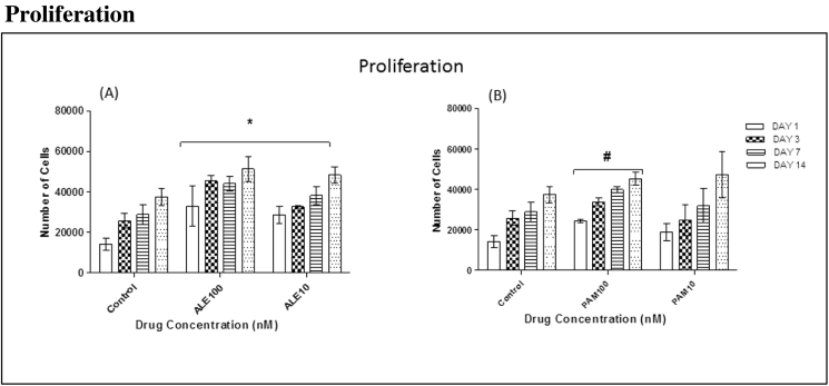
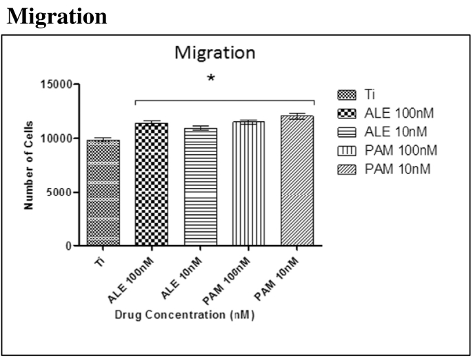
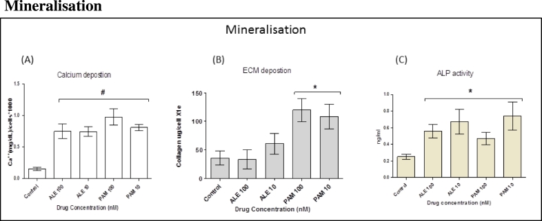
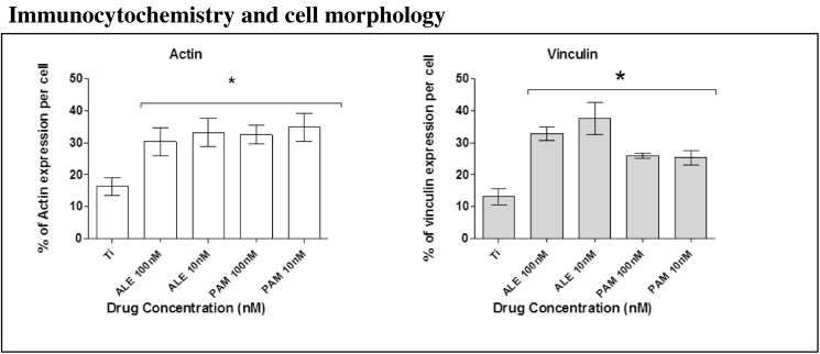
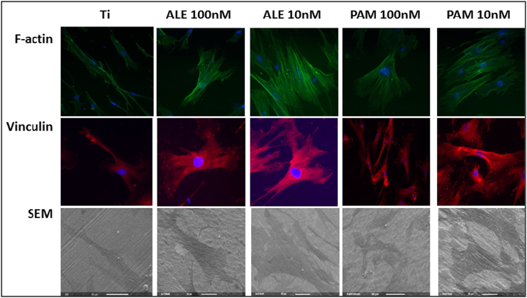
Since the 1980s, titanium (Ti) implants have been routinely used to replace missing teeth. This success is mainly due to the good biocompatibility of Ti and the phenomenon of osseointegration, with very early events at implant placement being important in determining good osseointegration. However, enhancing implant performance with coatings such as hydroxyapatite (HA) and calcium phosphate has proved largely unsuccessful. Human mesenchymal stem cells (hMSCs) are the first osteogenic cells to colonise implant surfaces and offer a target for enhancing osseointegration. We previously reported that small doses of bisphosphonate (BP) may play an integral role in enhancing hMSC proliferation and osteogenic differentiation. The aim of this study is to investigate whether small doses of bisphosphonates enhance proliferation and osteogenic differentiation of hMSCs on Ti surfaces, to enhance bone osseointegration and to accelerate wound healing around the implant surface. Our data suggests that treating cells with small doses of BP (100 nM & 10 nM) induces significant hMSC stimulation of osteogenic markers including calcium, collagen type I and ALP compared to control group on titanium surfaces (P < 0.05). In addition, cell proliferation and migration were significantly enhanced on titanium surfaces (P < 0.05).

Keywords: Bisphosphonates; Bone remodelling/regeneration; Human mesenchymal stem cells; Titanium.


Figures

Fig. 1
Fig. 1
The effects of low doses of ALE and PAM (100 nM and 10 nM) on hMSC proliferation on Ti surfaces were assessed at different time points. (A): All groups treated with ALE (100 nM and 10 nM) had shown significant cell proliferation when compared to the control group that was treated with GM only on day 1, 3, 7 and 14. (B): The group treated with 100 nM showed significant cell proliferation compared to the control group that treated with GM only on day 1, 3, 7 and 14 (values = mean ± SD; P < 0.05).
Fig. 2
Fig. 2
Significant hMSC migration was observed after 4.5 h incubation at 37 °C at 5% CO2. Significant differences were observed between the cells treated with the low dose of drugs and control group (OM only). Each column represents the mean ± SD.
Fig. 3
Fig. 3
Cells were divided into five groups with each group treated with different ALE and PAM concentration (100 nM and 10 nM). (A): At day 21, calcium deposition was analysed. The results showed that BPs significantly stimulated hMSC mineralisation following drug treatment for 21 days when compared to cells that had been treated with OM only. (B): At day 14, extracellular collagen formation was analysed. The results showed that PAM significantly stimulated hMSC to produce collagen following treatment with drugs for 14 days when compared to cells that had been treated with OM only. No significant changes were observed in cells treated with ALE when compared to the control. (C): ALP activity after 7 days incubation was significantly increased in all cells treated with the lower dose of BPs drug when compared to the control group treated with OM only. Each column represents the mean ± SD.
Fig. 4
Fig. 4
Actin and vinculin expression analysis after 24 h of culture in OM with and without BPs. Groups that been treated with low dose of ALE and PAM showed a significant increase in actin and vinculin expression compared to the control group that was treated with only OM. Each column represents the mean ± SD.
Fig. 5
Fig. 5
Fluorescence microscopy images showing the effect of low dose of drugs on cell organisation and spreading on Ti surfaces after 24 h. Actin expression (green images) appeared more abundant in cells treated with the ALE and PAM (100 nM and 10 nM). Likewise, vinculin expression (red images) appeared more abundant in treated cells. Images were taken using a × 40 objective. Scale bar = 50 μM. SEM images were found to correlate with fluorescent images.

References

    1. Abtahi J., Tengvall P., Aspenberg P. A bisphosphonate-coating improves the fixation of metal implants in human bone. A randomized trial of dental implants. Bone. 2012;50:1148–1151. - PubMed
    1. Alqhtani N.R., Meghji S., Brett P. International Association for Dental Research (IADR). IADR; 2014. The Effect of Bisphosphonates on hMSCs Proliferation and Osteogenic Differentiation.
    1. Baas J., Jakobsen T., Elmengaard B., Bechtold J.E., Soballe K. The effect of adding an equine bone matrix protein lyophilisate on fixation and osseointegration of HA-coated Ti implants. J. Biomed. Mater. Res. A. 2012;100:188–194. - PMC - PubMed
    1. Berruti A., Dogliotti L., Tucci M., Tarabuzzi R., Fontana D., Angeli A. Metabolic bone disease induced by prostate cancer: rationale for the use of bisphosphonates. J. Urol. 2001;166:2023–2031. - PubMed
    1. Chrcanovic B.R., Martins M.D., Wennerberg A. Immediate placement of implants into infected sites: a systematic review. Clin. Implant. Dent. Relat. Res. 2015;17(Suppl. 1):e1–e16. - PubMed

LinkOut - more resources