

Central activation of the A1 adenosine receptor in fed mice recapitulates only some of the attributes of daily torpor
- PMID: 28378088
- PMCID: PMC5493318
- DOI: 10.1007/s00360-017-1084-7
Central activation of the A1 adenosine receptor in fed mice recapitulates only some of the attributes of daily torpor
Abstract
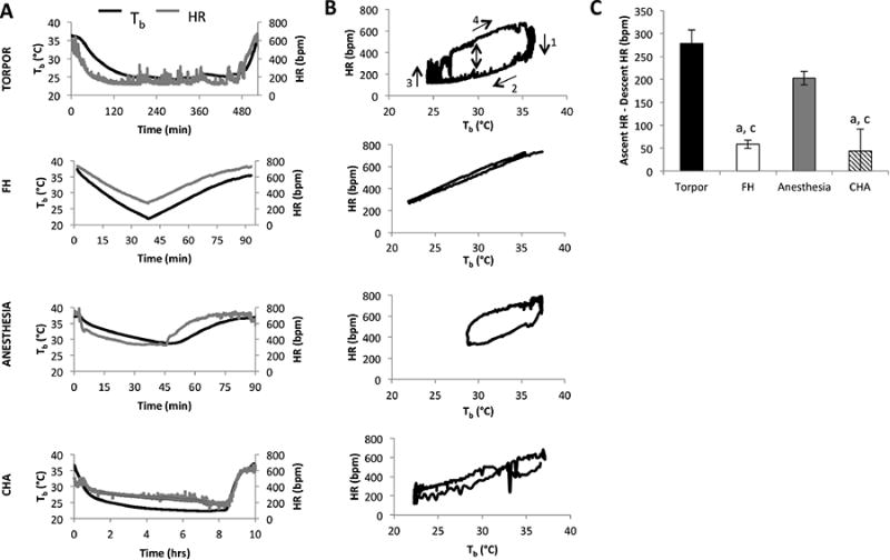
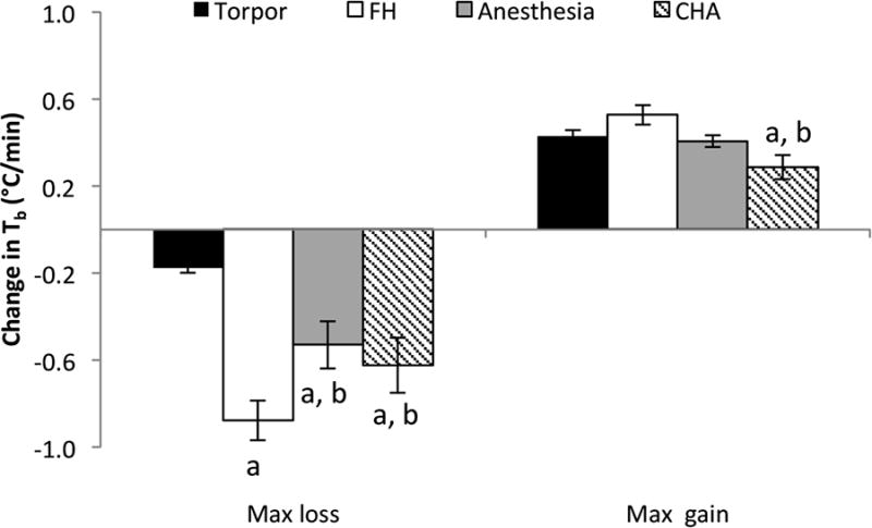
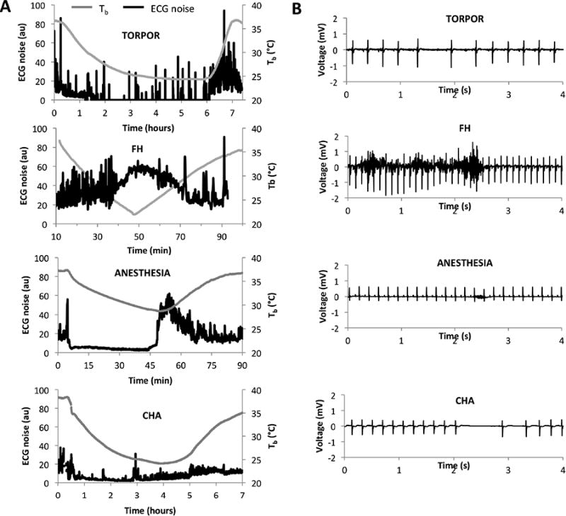
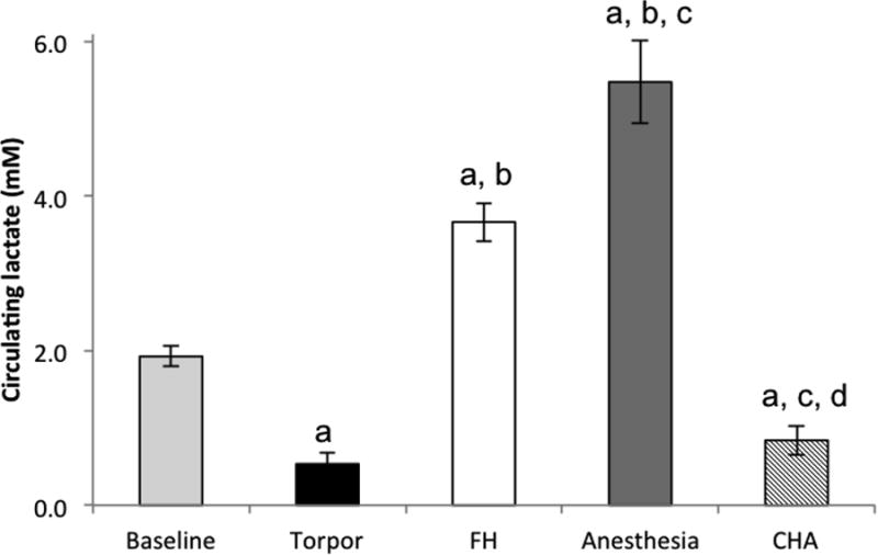
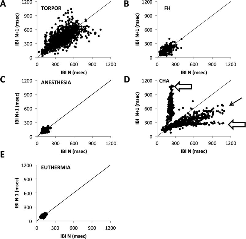
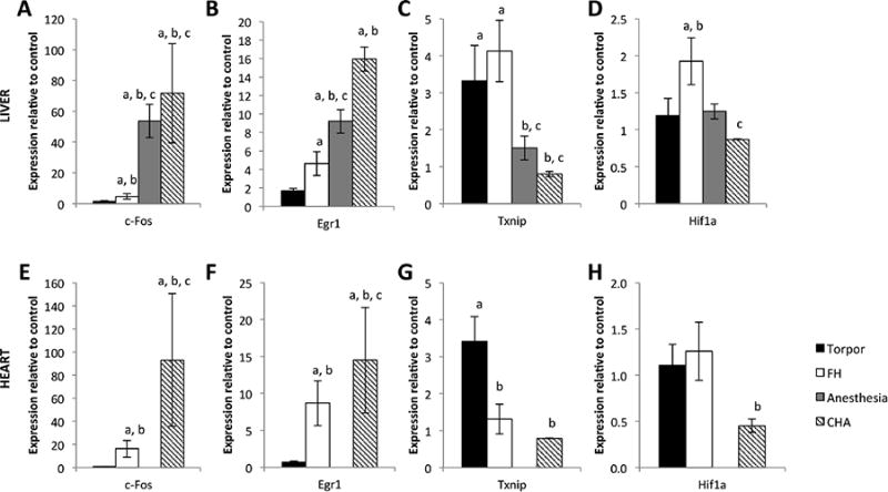
Mice enter bouts of daily torpor, drastically reducing metabolic rate, core body temperature (T b), and heart rate (HR), in response to reduced caloric intake. Because central adenosine activation has been shown to induce a torpor-like state in the arctic ground squirrel, and blocking the adenosine-1 (A1) receptor prevents daily torpor, we hypothesized that central activation of the A1 adenosine receptors would induce a bout of natural torpor in mice. To test the hypothesis, mice were subjected to four different hypothermia bouts: natural torpor, forced hypothermia (FH), isoflurane-anesthesia, and an intracerebroventricular injection of the selective A1 receptor agonist N6-cyclohexyladenosine (CHA). All conditions induced profound hypothermia. T b fell more rapidly in the FH, isoflurane-anesthesia, and CHA conditions compared to torpor, while mice treated with CHA recovered at half the rate of torpid mice. FH, isoflurane-anesthesia, and CHA-treated mice exhibited a diminished drop in HR during entry into hypothermia as compared to torpor. Mice in all conditions except CHA shivered while recovering from hypothermia, and only FH mice shivered substantially while entering hypothermia. Circulating lactate during the hypothermic bouts was not significantly different between the CHA and torpor conditions, both of which had lower than baseline lactate levels. Arrhythmias were largely absent in the FH and isoflurane-anesthesia conditions, while skipped beats were observed in natural torpor and periodic extended (>1 s) HR pauses in the CHA condition. Lastly, the hypothermic bouts showed distinct patterns of gene expression, with torpor characterized by elevated hepatic and cardiac Txnip expression and all other hypothermic states characterized by elevated c-Fos and Egr-1 expression. We conclude that CHA-induced hypothermia and natural torpor are largely different physiological states.
Keywords: Adenosine; Hibernation; Hypothermia; Targeted temperature management; Torpor.
Figures
Comment in
-
Central adenosine and daily torpor in mice.Temperature (Austin). 2017 Aug 2;4(4):350-352. doi: 10.1080/23328940.2017.1345713. eCollection 2017. Temperature (Austin). 2017. PMID: 29435475 Free PMC article. No abstract available.
Similar articles
-
Hypothermia in mouse is caused by adenosine A1 and A3 receptor agonists and AMP via three distinct mechanisms.Neuropharmacology. 2017 Mar 1;114:101-113. doi: 10.1016/j.neuropharm.2016.11.026. Epub 2016 Nov 30. Neuropharmacology. 2017. PMID: 27914963 Free PMC article.
-
Central activation of the A1 adenosine receptor (A1AR) induces a hypothermic, torpor-like state in the rat.J Neurosci. 2013 Sep 4;33(36):14512-25. doi: 10.1523/JNEUROSCI.1980-13.2013. J Neurosci. 2013. PMID: 24005302 Free PMC article.
-
Translating drug-induced hibernation to therapeutic hypothermia.ACS Chem Neurosci. 2015 Jun 17;6(6):899-904. doi: 10.1021/acschemneuro.5b00056. Epub 2015 Apr 8. ACS Chem Neurosci. 2015. PMID: 25812681 Free PMC article.
-
[Pharmacological aspects of mammalian hibernation: central thermoregulation factors in hibernation cycle].Nihon Yakurigaku Zasshi. 2000 Nov;116(5):304-12. doi: 10.1254/fpj.116.304. Nihon Yakurigaku Zasshi. 2000. PMID: 11215381 Review. Japanese.
-
Antipsychotic inductors of brain hypothermia and torpor-like states: perspectives of application.Psychopharmacology (Berl). 2017 Jan;234(2):173-184. doi: 10.1007/s00213-016-4496-2. Epub 2016 Dec 8. Psychopharmacology (Berl). 2017. PMID: 27933367 Review.
Cited by
-
Shallow metabolic depression and human spaceflight: a feasible first step.J Appl Physiol (1985). 2020 Mar 1;128(3):637-647. doi: 10.1152/japplphysiol.00725.2019. Epub 2020 Jan 30. J Appl Physiol (1985). 2020. PMID: 31999524 Free PMC article.
-
The relationship between fasting-induced torpor, sleep, and wakefulness in laboratory mice.Sleep. 2021 Sep 13;44(9):zsab093. doi: 10.1093/sleep/zsab093. Sleep. 2021. PMID: 33838033 Free PMC article.
-
Adenosine and P1 receptors: Key targets in the regulation of sleep, torpor, and hibernation.Front Pharmacol. 2023 Mar 10;14:1098976. doi: 10.3389/fphar.2023.1098976. eCollection 2023. Front Pharmacol. 2023. PMID: 36969831 Free PMC article. Review.
-
Estrogen-sensitive medial preoptic area neurons coordinate torpor in mice.Nat Commun. 2020 Dec 11;11(1):6378. doi: 10.1038/s41467-020-20050-1. Nat Commun. 2020. PMID: 33311503 Free PMC article.
-
Adenosine receptor A1 enhanced mitochondrial biogenesis and exerted neuroprotection after cerebral ischemia through PGC-1α.Exp Brain Res. 2023 Jun;241(6):1471-1488. doi: 10.1007/s00221-023-06613-w. Epub 2023 Apr 20. Exp Brain Res. 2023. PMID: 37081178
References
-
- Ahmad FU, Wang MY, Levi AD. Hypothermia for acute spinal cord injury–a review. World Neurosurg. 2014;82(1-2):207–214. - PubMed
-
- Bouma HR, Verhaag EM, Otis JP, Heldmaier G, Swoap SJ, Strijkstra AM, Henning RH, Carey HV. Induction of torpor: Mimicking natural metabolic suppression for biomedical applications. J Cell Physiol 2011 - PubMed
MeSH terms
Substances
Grants and funding
LinkOut - more resources
Full Text Sources
Other Literature Sources
Medical
Miscellaneous
