

Rapid prototyping technology and its application in bone tissue engineering
- PMID: 28378568
- PMCID: PMC5394095
- DOI: 10.1631/jzus.B1600118
Rapid prototyping technology and its application in bone tissue engineering
Abstract
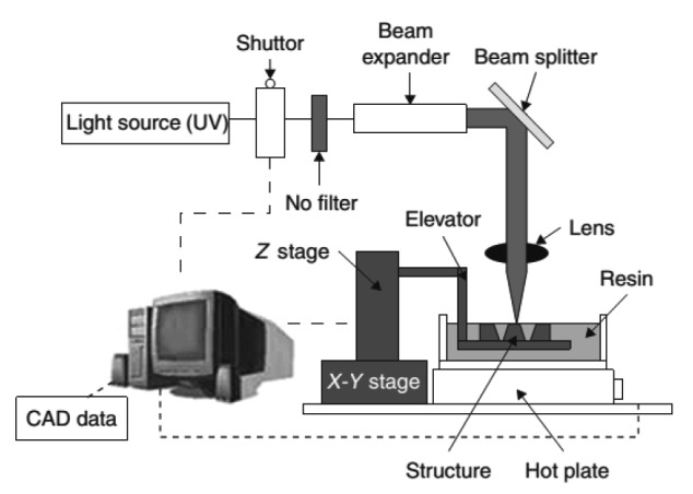
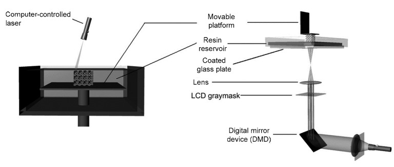
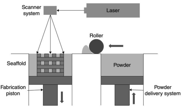
Bone defects arising from a variety of reasons cannot be treated effectively without bone tissue reconstruction. Autografts and allografts have been used in clinical application for some time, but they have disadvantages. With the inherent drawback in the precision and reproducibility of conventional scaffold fabrication techniques, the results of bone surgery may not be ideal. This is despite the introduction of bone tissue engineering which provides a powerful approach for bone repair. Rapid prototyping technologies have emerged as an alternative and have been widely used in bone tissue engineering, enhancing bone tissue regeneration in terms of mechanical strength, pore geometry, and bioactive factors, and overcoming some of the disadvantages of conventional technologies. This review focuses on the basic principles and characteristics of various fabrication technologies, such as stereolithography, selective laser sintering, and fused deposition modeling, and reviews the application of rapid prototyping techniques to scaffolds for bone tissue engineering. In the near future, the use of scaffolds for bone tissue engineering prepared by rapid prototyping technology might be an effective therapeutic strategy for bone defects.
Keywords: Rapid prototyping; Bone tissue engineering; Scaffolds.
Conflict of interest statement
This article does not contain any studies with human or animal subjects performed by any of the authors.
Figures
References
-
- Arcaute K, Mann B, Wicker R. Stereolithography of spatially controlled multi-material bioactive poly(ethylene glycol) scaffolds. Acta Biomater. 2010;6(3):1047–1054. (Available from: http://dx.doi.org/10.1016/j.actbio.2009.08.017) - DOI - PubMed
-
- Bose S, Roy M, Bandyopadhyay A. Recent advances in bone tissue engineering scaffolds. Trends Biotechnol. 2012;30(10):546–554. (Available from: http://dx.doi.org/10.1016/j.tibtech.2012.07.005) - DOI - PMC - PubMed
-
- Bose S, Vahabzadeh S, Bandyopadhyay A. Bone tissue engineering using 3D printing. Mater Today. 2013;16(12):496–504. (Available from: http://dx.doi.org/10.1016/j.mattod.2013.11.017) - DOI
-
- Brie J, Chartier T, Chaput C, et al. A new custom made bioceramic implant for the repair of large and complex craniofacial bone defects. J Cranio Maxill Surg. 2013;41(5):403–407. (Available from: http://dx.doi.org/10.1016/j.jcms.2012.11.005) - DOI - PubMed
-
- Calori GM, Mazza E, Colombo M, et al. The use of bone-graft substitutes in large bone defects: any specific needs? Injury. 2011;42(Suppl. 2):S56–S63. (Available from: http://dx.doi.org/10.1016/j.injury.2011.06.011) - DOI - PubMed
Publication types
MeSH terms
Substances
LinkOut - more resources
Full Text Sources
Other Literature Sources
