. 2017 Apr 5:7:45319.
doi: 10.1038/srep45319.

Siglec-7 restores β-cell function and survival and reduces inflammation in pancreatic islets from patients with diabetes

Affiliations

Siglec-7 restores β-cell function and survival and reduces inflammation in pancreatic islets from patients with diabetes

Gitanjali Dharmadhikari et al. Sci Rep. .

Abstract

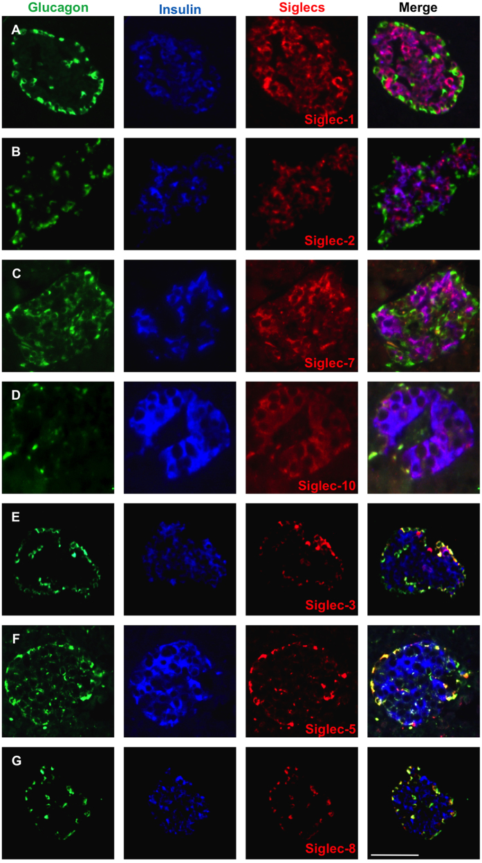
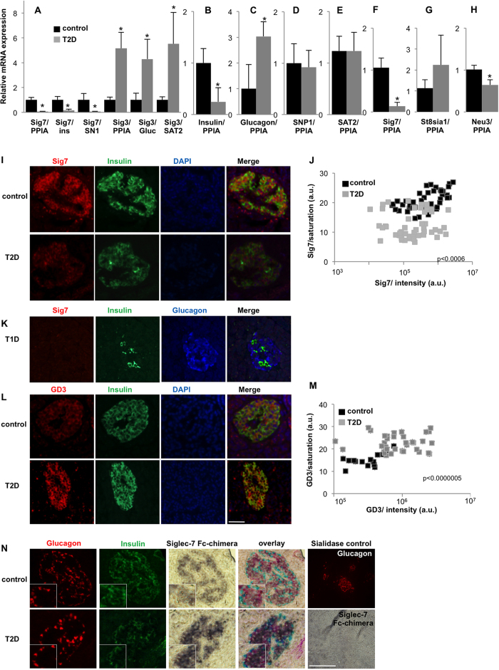
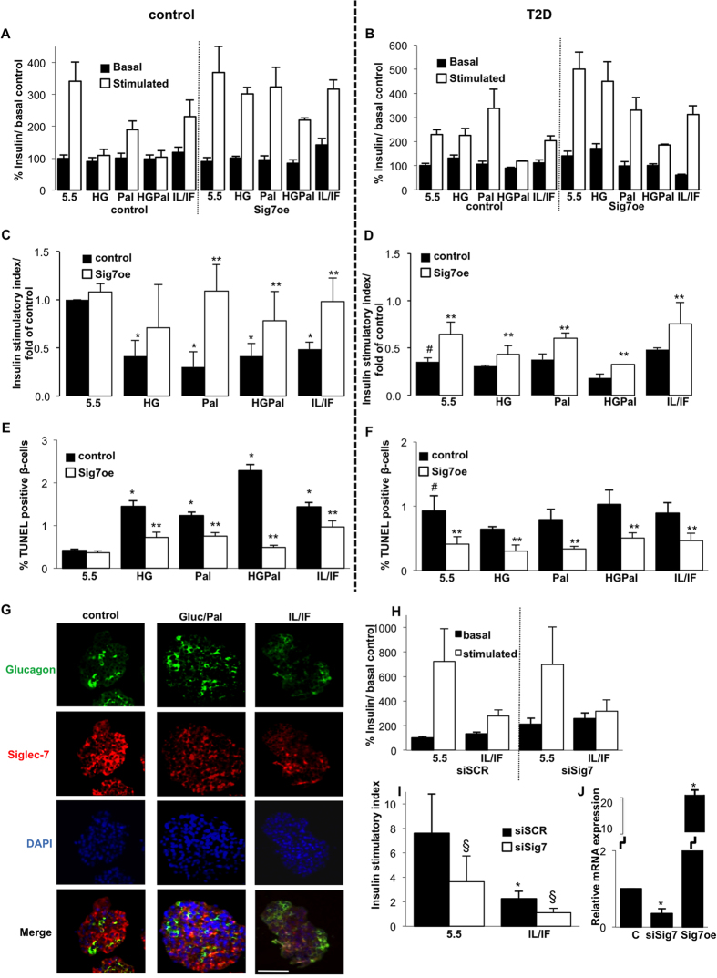
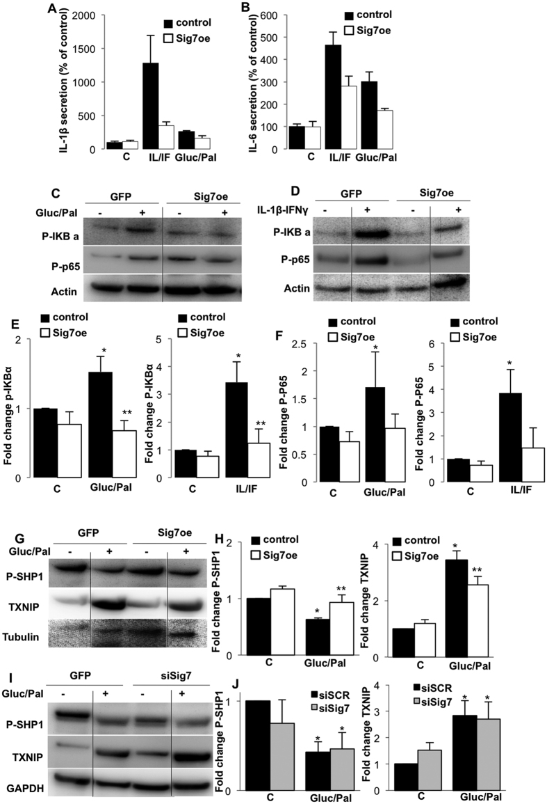
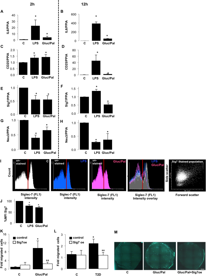
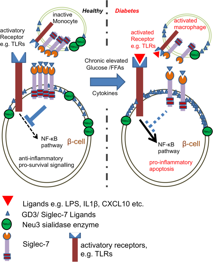
Chronic inflammation plays a key role in both type 1 and type 2 diabetes. Cytokine and chemokine production within the islets in a diabetic milieu results in β-cell failure and diabetes progression. Identification of targets, which both prevent macrophage activation and infiltration into islets and restore β-cell functionality is essential for effective diabetes therapy. We report that certain Sialic-acid-binding immunoglobulin-like-lectins (siglecs) are expressed in human pancreatic islets in a cell-type specific manner. Siglec-7 was expressed on β-cells and down-regulated in type 1 and type 2 diabetes and in infiltrating activated immune cells. Over-expression of Siglec-7 in diabetic islets reduced cytokines, prevented β-cell dysfunction and apoptosis and reduced recruiting of migrating monocytes. Our data suggest that restoration of human Siglec-7 expression may be a novel therapeutic strategy targeted to both inhibition of immune activation and preservation of β-cell function and survival.


Conflict of interest statement

The authors declare no competing financial interests.

Figures

Figure 1
Figure 1. Siglecs are differentially expressed in the human Islets of Langerhans.
Triple immunostaining for insulin (blue), glucagon (green) and siglecs (red) was carried out on human pancreatic sections obtained at autopsy from non-diabetic individuals. (A) Siglecs-1, (B) -2, (C) -7and (D) -10 were expressed in β-cells. (E) Siglecs-3, (F) -5, (G) -8 were expressed solely in α-cells. Representative analyses from 5 pancreases from age and weight-matched patients with T2D and 5 controls are shown. Bar is 100 μm.
Figure 2
Figure 2. Siglec-7 and -3 are reciprocally regulated in type 2 diabetes.
Semi quantitative real time PCR analysis was performed on cDNAs obtained from autopsy pancreases from non-diabetic (n = 9) and individuals with T2D (n = 5), the latter all with documented fasting plasma glucose >150 mg/dl. (A) Siglec-7 expression was normalized on cyclophilin (PPIA), insulin (ins) and SN1; whereas Siglec-3 expression was normalized on cyclophilin, glucagon (gluc) and SAT2. (B–E) Insulin, Glucagon, SN1 and SAT2 were normalized on cyclophilin. (F–H) Real time PCR analysis of freshly isolated islets of patients with T2D (n = 5) were compared to that of non-diabetic individuals (n = 3) of (F) Siglec-7, (G) St8Sia1 and (H) Neu3 (I,J). Immunohistochemical analysis was carried out on human pancreatic sections obtained at autopsy of non-diabetic controls and patients with T2D for (I,J) Siglec-7, (L) GD3. (J,M) staining saturation and intensity were quantified using Photoshop; each data point represents saturation and intensity of the protein signal of islets from an average of 42 islets from 3 donors, respectively. (K) Analysis of Siglec-7 and insulin in the pancreas of a patient with T1D with remaining insulin+β-cells. (N) Bright field staining using Siglec-7 Fc-chimeras along with glucagon (red) and insulin (green); along with control slides treated with sialidase treatment. Bar is 100 μm.
Figure 3
Figure 3. Siglec-7 over-expression improves β-cell survival and function.
Freshly isolated human islets of non-diabetic individuals as well as from patients with T2D were cultured on extracellular matrix-coated dishes and exposed to elevated glucose concentrations (22.2 or 33.3 mM; HG-both had the same effect and thus results were combined) with or without palmitate (HGPal), palmitate alone (Pal) or the cytokine mixture IL-1β (2 ng/ml) and IFNγ (1,000 U/ml) (IL/IF) for 72 h with or without over-expression by lipofectamine-mediated Siglec-7 plasmid transfection. Glucose stimulated insulin secretion assays were performed after the 72 h culture period. (A,B) Basal (2.8 mM) and glucose stimulated (16.7 mM) insulin secretion was expressed as percent change of control condition basal insulin levels. (C,D) Stimulatory index denotes the amount of glucose stimulated (16.7 mM glucose) divided by the amount of basal insulin secretion. Fold changes in stimulatory indices of treated islets were plotted, compared to stimulatory index of control islets. (E,F) Apoptosis was analyzed by the TUNEL assay in dishes. Islets were triple-stained for insulin and counterstained for DAPI (not shown). Results are means ± SE of the percentage of TUNEL-positive β-cells. The average number of β-cells counted was 8124 for each treatment group in 3–4 separate experiments from 3 separate dishes per treatment from 3–4 different organ donors. (G) Isolated human islets were treated with 22.2 mM glucose and 0.5 mM palmitate; or the cytokine mixture IL/IF, followed by immunohistochemical analysis of paraffin-embedded islet sections. Representative images show glucagon (green), Siglec-7 (red) and DAPI (blue). (H,I) Human islets were transfected with 100 nM siRNA to Siglec-7 or scrambled control (siSCR) and treated with the cytokine mixture IL/IF for 72 h. (H) Basal (2.8 mM) and glucose stimulated (16.7 mM) insulin secretion and (I) stimulatory index were analyzed as above. (J) Siglec mRNA expression in islets transfected with siRNA to Siglec-7 or Siglec-7 plasmid DNA. *p < 0.05 to 5.5 mM glucose non-treated control islets, **p < 0.05 Siglec-7 vs. LacZ transfected islets under same diabetic stimuli, #p < 0.05 to 5.5 mM glucose treated LacZ transfected non-diabetic control islets. §p < 0.05 to scramble siRNA treated islets under the same treatment. Data are shown as mean ± SE. Experiments were performed in triplicates, respectively from at least 3–5 independent experiments per condition from 3–5 human islet donors. Bar is 100 μm.
Figure 4
Figure 4. Siglec-7 inhibits NF-κB activation and cytokine secretion by pancreatic islets.
Human pancreatic islets were cultured on extracellular matrix-coated dishes and exposed to elevated glucose (22.2 mM) and palmitate (Gluc/Pal) or the cytokine mixture IL-1β (2 ng/ml) and IFNγ (1,000 U/ml) (IL/IF) for 72 h with or without over-expression of lipofectamine-mediated Siglec-7 plasmid transfection. (A,B) The cytokine profiles of the supernatants of transfected and treated islets were assessed using protein array ELISAs for IL-1β and IL-6; absolute values at control are 4.2 + /−4.75 pg/ml IL-1β and 458 + /−51 pg/ml IL-6. (C–F) Western blot analysis was performed after Siglec-7 over-expression in islets and 72 h treatment with 22.2 mM glucose and palmitate or IL-1β and IFNγ; and analyzed for P-p65, P-IKBα and actin. (G–J) Western blot analysis of P-SHP1 and TXNIP from isolated human islets overexpressing Siglec-7 or after Siglec-7 silencing and exposure to 22.2 mM glucose and palmitate for 72 h. (E,F,H,J) Densitometry analysis of bands normalized on housekeeping proteins and plotted as fold change of islets at control condition. (A,B) are means of 2 independent experiments from 2 different organ donors from 6 dishes per treatment condition. All blots are representative of 3–5 independent experiments and densitometry are means of 3–5 independent experiments from 3–5 different organ donors. Lanes were run on the same gel but were noncontiguous. Full blots are shown in the Suppl. Fig. section “Full blots”. *p < 0.05 to 5.5 mM glucose non-treated control islets, **p < 0.05 Siglec-7 vs. LacZ transfected islets under same diabetic stimuli.
Figure 5
Figure 5. Immune cell migration into inflamed islets is inhibited by Siglec-7.
PBMCs purified from buffy coats of blood donors (n = 6) were treated with lipopolysaccharide (LPS) or elevated glucose and palmitate (Gluc/Pal) for (A,C,E,G) 2 h or (B,D,F,H) 12 h. Real time PCR analysis of these treated cells was carried out for (A,B) IL-6, (C,D) CD25, (E,F) Siglec-7 and (G,H) Neu-3. (I) Cell surface expression of Siglec-7 in the treated PBMCs was determined using flow cytometry. Histograms for intensity of Siglec-7 (FL1 filter) was plotted and overlayed to observe the effect of these treatments. (J) Histograms were quantified and Siglec-7 expression was plotted as % mean fluorescent intensity as compared to untreated control fraction. The migration of leukocytes (n = 3 buffy coat donors) in response to conditioned media obtained from transfected and treated islets (n = 3 separate dishes from 3 independent experiments from 3 donors), was quantified after 4 h using an in vitro migration assay. (K) The fold induction of migration as compared to untreated control islet supernatants was plotted. (L) Migration of mononuclear cells (n = 3) with respect to cultured islets from donors with T2D (n = 3), with or without Siglec-7 over-expression, was plotted as fold change of migrated cells compared to untreated control islets of a non-diabetic individual. (M) The images are representative of fluorescent microscopic analysis of live cells migrating through membranes observed in green (shown in (K)). For PBMC treatments; *p < 0.05 to 11.1 mM glucose treated monocyte fraction. For migration assay, *p < 0.05 to monocyte fraction treated with 5.5 mM glucose, LacZ transfected control islets, **p < 0.05 to monocyte fraction treated with Gluc/Pal, LacZ transfected control islets.
Figure 6
Figure 6. Our view on the deleterious loss of Siglec-7 signals under diabetogenic conditions.
In healthy individuals, Siglec-7 helps to maintain a pro-survival anti-inflammatory signaling in monocytes as well as in β-cells. The membrane-associated sialic acid-cleaving enzyme sialidase Neu3 unmasks Siglec-7 residues and makes them free for binding and activation and thus enhances Siglec-7 inhibitory downstream cell protection events (e.g. NFκB inhibition). In diabetes, chronically elevated glucose along with palmitate and cytokines cause loss of Siglec-7 in these cells. The simultaneous loss in the sialidase Neu3 and the increase in endogenous Siglec-7 ligands block siglec downstream signals. This leads to triggering of apoptotic and pro-inflammatory signals, activation of macrophages and ultimately to β-cell death.

References

    1. Donath M. Y., Storling J., Maedler K. & Mandrup-Poulsen T. Inflammatory mediators and islet beta-cell failure: a link between type 1 and type 2 diabetes. J. Mol. Med. 81, 455–470 (2003). - PubMed
    1. Tisch R. & McDevitt H. Insulin-dependent diabetes mellitus. Cell 85, 291–297 (1996). - PubMed
    1. Kurrer M. O., Pakala S. V., Hanson H. L. & Katz J. D. Beta cell apoptosis in T cell-mediated autoimmune diabetes. Proc. Natl. Acad. Sci. USA 94, 213–218 (1997). - PMC - PubMed
    1. Mokdad A. H. et al.. Prevalence of obesity, diabetes, and obesity-related health risk factors, 2001. JAMA 289, 76–79 (2003). - PubMed
    1. Neels J. G. & Olefsky J. M. Inflamed fat: what starts the fire? J Clin Invest 116, 33–35 (2006). - PMC - PubMed

Publication types

MeSH terms