. 2017 Jun;25(6):771-774.
doi: 10.1038/ejhg.2017.45. Epub 2017 Apr 5.

Upstream SLC2A1 translation initiation causes GLUT1 deficiency syndrome

Affiliations

Upstream SLC2A1 translation initiation causes GLUT1 deficiency syndrome

Michèl A Willemsen et al. Eur J Hum Genet. 2017 Jun.

Abstract

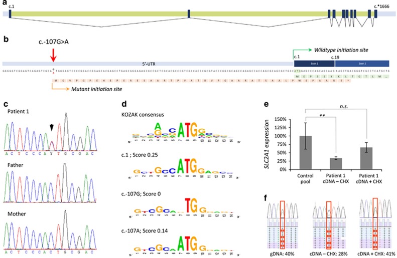
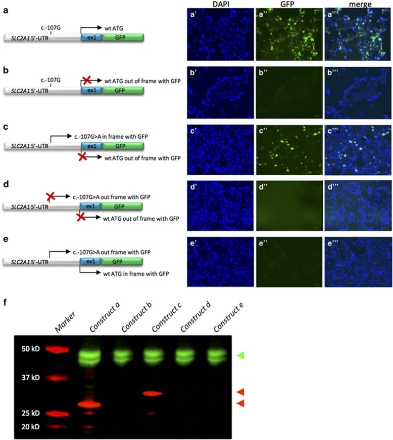
Glucose transporter type 1 deficiency syndrome (GLUT1DS) is a neurometabolic disorder with a complex phenotypic spectrum but simple biomarkers in cerebrospinal fluid. The disorder is caused by impaired glucose transport into the brain resulting from variants in SCL2A1. In 10% of GLUT1DS patients, a genetic diagnosis can not be made. Using whole-genome sequencing, we identified a de novo 5'-UTR variant in SLC2A1, generating a novel translation initiation codon, severely compromising SLC2A1 function. This finding expands our understanding of the disease mechanisms underlying GLUT1DS and encourages further in-depth analysis of SLC2A1 non-coding regions in patients without variants in the coding region.


Conflict of interest statement

JK has received speakers honoraria and travel costs from Nutricia GmbH, Eerlangen, Germany and Vitaflo Pharma GmbH, Bad Homburg, Germany. The remaining authors declare no conflict of interest.

Figures

Figure 1
Figure 1
Molecular characterization and expression analysis of the SLC2A1 c.-107G>A variant. (a) Schematic depiction of the SLC2A1 exons (black boxes), introns (green bars), and UTRs (gray bars) with splicing information (dotted lines) based on NM_006516.2. (b) The c.-107G>A variant results in a potential novel translation initiation site. The predicted protein effect is indicated in orange, whereas the wild-type protein (partial) is shown in green. The mutant protein is predicted to result in a premature termination codon in exon 2 (*). (c) Sanger sequencing validation of the c.-107G>A variant in patient 1 and her parents, indicating de novo occurrence of the variant. (d) Evaluation of the sequence context surrounding c.107G for the presence of a KOZAK sequence. Top panel shows the consensus sequence, with the relative height of the nucleotides representing the frequency observed at the specified position. Second panel shows the wild-type SLC2A1 c.1 KOZAK sequence. Third panel shows the predication for the wild-type c.107G sequence and its absence of a KOZAK sequence. Lower panel shows the prediction for c.107 A with the presence of a KOZAK sequence. Scores represent reliability of the KOZAK prediction. (e) Relative SLC2A1 mRNA expression in patient 1, indicating a significantly reduced expression (cDNA – CHX) that is partially increased by blocking of nonsense-mediated decay with cycloheximide (cDNA+CHX); **P<0.01. (f) Nonsense-mediated decay of the SLC2A1 transcript bearing the c.-107G>A variant. Patient's fibroblasts were cultured in the absence (−CHX) or presence (+CHX) of cycloheximide, and both genomic (gDNA) and mRNA were isolated. Copy-DNA (cDNA) was made from the mRNA samples, and both gDNA and cDNA were subjected to semi-quantitative ion-torrent sequencing (>40 000 reads per sample) after PCR. The percentage of reads containing the c.-107G>A variant is given. NS, not significant.
Figure 2
Figure 2
Functional characterization of SLC2A1 c.-107G>A. HEK293T cells were transfected with constructs of the 5′-UTR and exon 1 of SLC2A1 fused to GFP. Left: cartoons of the constructs used. The positions and possible use (arrow) of the wild-type initiation codon (ae) and the novel initiation codon introduced by the c.-107G>A variant (ce) are indicated. If no expression is anticipated, the arrow is crossed out (X). Right: representative images of cells transfected with the respective constructs with signals for nuclei by DAPI staining (blue) or green fluorescent protein (green). (f) Western blot of the SLC2A1-GFP fusion proteins created by the constructs in HEK293T cells. An antibody directed against tubulin was used as control (in green, top arrow head), whereas an antibody against GFP was used to detect the SLC2A1-GFP fusion (in red). The fusion proteins resulting from usage of the novel initiation site or the wild-type initiation codon are 32 kD (upper red arrow head) and 29 kD (lower red arrow head) in size, respectively.

References

    1. De Vivo DC, Trifiletti RR, Jacobson RI et al: Defective glucose transport across the blood-brain barrier as a cause of persistent hypoglycorrhachia, seizures, and developmental delay. N Engl J Med 1991; 325: 703–709. - PubMed
    1. Seidner G, Alvarez MG, Yeh JI et al: GLUT-1 deficiency syndrome caused by haploinsufficiency of the blood-brain barrier hexose carrier. Nat Genet 1998; 18: 188–191. - PubMed
    1. Leen WG, Klepper J, Verbeek MM et al: Glucose transporter-1 deficiency syndrome: the expanding clinical and genetic spectrum of a treatable disorder. Brain 2010; 133: 655–670. - PubMed
    1. Hully M, Vuillaumier-Barrot S, Le Bizec C et al: From splitting GLUT1 deficiency syndromes to overlapping phenotypes. Eur J Med Genet 2015; 58: 443–454. - PubMed
    1. Liu YC, Lee JW, Bellows ST et al: Evaluation of non-coding variation in GLUT1 deficiency. Dev Med Child Neurol 2016; 58: 1295–1302. - PubMed

MeSH terms

Supplementary concepts