. 2017 May 8;19(5):299-307.
doi: 10.1021/acscombsci.6b00182. Epub 2017 Apr 13.

Combinatorial Library Screening with Liposomes for Discovery of Membrane Active Peptides

Affiliations

Combinatorial Library Screening with Liposomes for Discovery of Membrane Active Peptides

Randy P Carney et al. ACS Comb Sci. .

Abstract

Membrane active peptides (MAPs) represent a class of short biomolecules that have shown great promise in facilitating intracellular delivery without disrupting cellular plasma membranes. Yet their clinical application has been stalled by numerous factors: off-target delivery, a requirement for high local concentration near cells of interest, degradation en route to the target site, and in the case of cell-penetrating peptides, eventual entrapment in endolysosomal compartments. The current method of deriving MAPs from naturally occurring proteins has restricted the discovery of new peptides that may overcome these limitations. Here, we describe a new branch of assays featuring high-throughput functional screening capable of discovering new peptides with tailored cell uptake and endosomal escape capabilities. The one-bead-one-compound (OBOC) combinatorial method is used to screen libraries containing millions of potential MAPs for binding to synthetic liposomes, which can be adapted to mimic various aspects of limiting membranes. By incorporating unnatural and d-amino acids in the library, in addition to varying buffer conditions and liposome compositions, we have identified several new highly potent MAPs that improve on current standards and introduce motifs that were previously unknown or considered unsuitable. Since small variations in pH and lipid composition can be controlled during screening, peptides discovered using this methodology could aid researchers building drug delivery platforms with unique requirements, such as targeted intracellular localization.

Keywords: drug delivery platforms; endosomal escape capabilities; high-throughput; liposomes; membrane-active proteins; one-bead-one-compound.


Conflict of interest statement

The authors declare no competing financial interests.

Figures
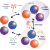
Figure 1
Figure 1
Sequential OBOC Screening Scheme. Beads from an OBOC combinatorial peptide library are immobilized on a planar polystyrene surface before incubation with fluorescently-labeled giant unilamellar vesicles (GUVs). The library can be washed with ethanol to remove bound GUVs for quick re-screening of the same beads under various conditions such as GUV composition and pH. More than 200,000 beads can be scanned within 20 minutes by automating a confocal microscopy tile-scan program. Beads are individually ranked for changes in fluorescence intensity over time due to binding of fluorescently-labeled GUVs, and subsequently compared across screening conditions.
Figure 2
Figure 2
Detection and selection scheme. (a) CLSM overlay of transmitted light (polymer beads) and red fluorescence channel (GUVs) during a typical screening. Beads that bind many GUVs (right) are selected as peptides of interest while non-binding beads (left) are ignored. (b) Red fluorescence channel CLSM image with GUV curvature evident. (c) Each bead’s autofluorescence signal (green channel) is used to spatially map out hundreds of thousands of beads in a single polystyrene dish. (d) An automated program can identify specific beads (e.g. bead 1414 pictured in (c)) based on discrimination of change in fluorescence over time. (e) The top several beads can be quickly picked and sequenced for further investigation.
Figure 3
Figure 3
Screening results and peptide library components. (a) Peptide sequences for five positive hits (termed bLips1–5) isolated after three screening rounds for binding simple fluorophore-labeled zwitterionic GUVs. (b) Peptide sequences identified with a more advanced screening methodology, which tracks each bead’s binding to either plasma membrane type or endosomal type GUVs at appropriate pH. (c) Amino acid abbreviations and corresponding side-chain functional groups comprising the combinatorial library screened in this study. Unnatural residues are blue.
Figure 4
Figure 4
In vitro uptake of bLips1–5 measured by confocal microscopy. (a) Flow cytometry measurement of mean fluorescence intensity (MFI) for A549 cells after incubation with FITC-labeled bLips for 3 h. (b) Left : CLSM fluorescence image illustrating TAMRA-conjugated bLip5 uptake in A549 lung cancer cells (blue channel-DAPI nuclear stain) Right: Higher magnification red fluorescence and transmitted light overlay CLSM image showing cellular localization of bLip5 in ES2 ovarian cancer cells.
Figure 5
Figure 5
(a) CLSM overlay of brightfield (cells) blue fluorescence channel (nuclei stain), and red fluorescence channel (TAMRA dye) following cell uptake of either bLip5-TAMRA (left) or R9-TAMRA (right). (b) Representative CLSM images were analyzed for fluorescence intensity normalized by cell area. The bLip5-TAMRA intensity is graphed as percent fluorescent intensity compared to R9-TAMRA. Error bars represent one standard deviation.
Figure 6
Figure 6
Toxicity of bLips measured by flow cytometry. (a) Representative flow cytometry raw fluorescence traces following propidium iodide addition for (i) A549 control cells (no peptide added), and A549 cells incubated with (ii) 30 µM R9, and (iii) 30 µM bLip1. (b) Percentage of viable A549 cells based on PI viability assay for R9 and the five peptide bLips at increasing concentration of 5, 30, and 100 µM (roman numerals indicate the data set referenced in (a)). The bLips show very little cytotoxicity, even at high concentration. C stands for control.
Figure 7
Figure 7
In vitro uptake of bLips6–9 measured by confocal microscopy. CLSM fluorescence image illustrating FITC-conjugated bLips6–9 uptake in A549 lung cancer cells (blue channel-DAPI nuclear stain) after 1 h incubation. bLips6–9 (identified to bind PM-GUVs at pH = 7.4) primarily showed punctate fluorescence indicative of endocytosis, while bLip 9 (binding only LE-GUVs at pH = 5.5) did not exhibit cell penetration.
Figure 8
Figure 8
In vitro GFP knockdown for peptides binding to LE-GUVs at pH = 5.5. CLSM micrographs were used to quantify GFP expression in U87-MG brain tumor cells, 72 h post treatment. For each case, several representative images were analyzed for fluorescence intensity (normalized by cell area). The average intensities are graphed as percent GFP expression compared to control (no treatment). Error bars represent one standard deviation. The combination of GFP-antisense-siRNA and Lipofectamine resulted in significant decrease in GFP expression (α = 0.05) compared to control or Lipofectamine only (LF only). When bLip9 was co-incubated at 20 µM with siRNA/Lipofectamine complexes, GFP expression was further decreased. At 20 µM, bLips7 and 8 did not significantly decrease GFP expression.

References

    1. Lehner R, Wang X, Marsch S, Hunziker P. Intelligent nanomaterials for medicine: Carrier platforms and targeting strategies in the context of clinical application. Nanomed-Nanotechnol. 2013;9:742–757. - PubMed
    1. Liang W, W Lam JK. In: Molecular Regulation of Endocytosis. Ceresa B, editor. Chapter 17 InTech; 2012.
    1. Hillaireau H, Couvreur P. Nanocarriers’ entry into the cell: relevance to drug delivery. Cell. Mol. Life Sci. 2009:2873–2896. - PMC - PubMed
    1. Last NB, Schlamadinger DE, Miranker AD. A common landscape for membrane-active peptides. Protein Sci. 2013;22:870–882. - PMC - PubMed
    1. Gautam A, Singh H, Tyagi A, Chaudhary K, Kumar R, Kapoor P, Raghava GPS. CPPsite: a curated database of cell penetrating peptides. Database. 2012;2012:bas015–bas015. - PMC - PubMed

Publication types