Performance of apparent diffusion coefficient of medial and lateral rectus muscles in Graves' orbitopathy
- PMID: 28379055
- PMCID: PMC5480799
- DOI: 10.1177/1971400917691993
Performance of apparent diffusion coefficient of medial and lateral rectus muscles in Graves' orbitopathy
Abstract
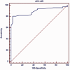
Objective The purpose of this study was to determine the performance of the apparent diffusion coefficient in the detection of involvement of the medial and lateral rectus muscles in patients with Graves' orbitopathy. Methods and materials This prospective study was conducted on 33 consecutive patients (16 males, 17 females with a mean age of 36 years) with Graves' orbitopathy and 18 age- and sex-matched volunteers. The patients and volunteers underwent diffusion-weighted magnetic resonance imaging of the orbit in the axial plane using echo-planar imaging. The apparent diffusion coefficient of the medial and lateral rectus muscles was calculated. Results The medial rectus muscle was more affected than the lateral rectus muscle. The mean apparent diffusion coefficient value of the medial and lateral rectus muscles was 1.81 ± 0.19 and 1.72 ± 0.07 × 10⁻³ mm2/s in patients with Graves' orbitopathy and 1.59 ± 0.06 and 1.51 ± 0.06 × 10⁻³ mm2/s in volunteers, respectively. There was a significant difference in apparent diffusion coefficient values of the medial and lateral rectus muscles between patients with Graves' orbitopathy and volunteers ( p = 0.001). The classification performance as measured with area under the receiver operator characteristic curve was 0.89 (95% confidence interval: 0.732-0.904). The best performing threshold of the apparent diffusion coefficient value of the medial rectus muscle was 1.69 × 10⁻³ mm2/s and associated efficiency was 86%, sensitivity was 97%, and specificity was 97%. Conclusion We concluded that the apparent diffusion coefficient of the medial rectus muscle can be used for diagnosis of Graves' orbitopathy.
Keywords: Diffusion; Graves; magnetic resonance imaging; orbit.
Figures


References
-
- Cockerham KP, Chan SS. Thyroid eye disease. Neurol Clin 2010; 28: 729–755. - PubMed
-
- Bahn RS. Current insights into the pathogenesis of Graves' ophthalmopathy. Horm Metab Res 2015; 47: 773–778. - PubMed
-
- Shan SJ, Douglas RS. The pathophysiology of thyroid eye disease. J Neuroophthalmol 2014; 34: 177–185. - PubMed
MeSH terms
LinkOut - more resources
Full Text Sources
Other Literature Sources

