. 2017;77(1):18-30.
doi: 10.21307/ane-2017-033.

Energy-dense diet triggers changes in gut microbiota, reorganization of gut‑brain vagal communication and increases body fat accumulation

Affiliations

Energy-dense diet triggers changes in gut microbiota, reorganization of gut‑brain vagal communication and increases body fat accumulation

Alexandra C Vaughn et al. Acta Neurobiol Exp (Wars). 2017.

Abstract

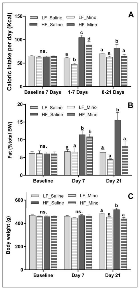
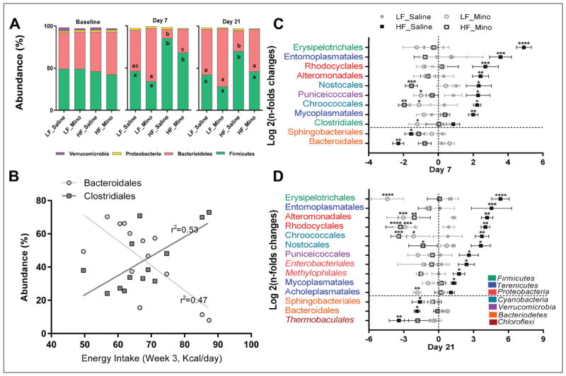
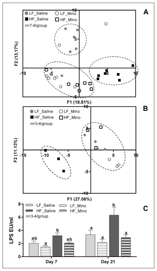
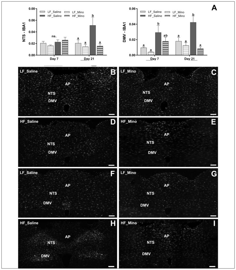
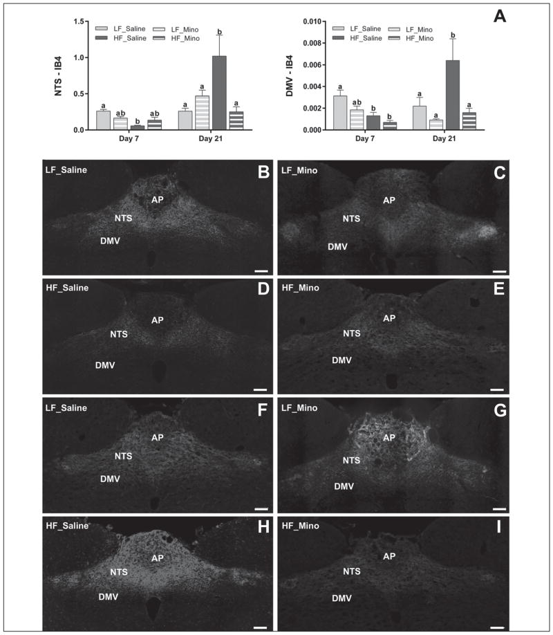
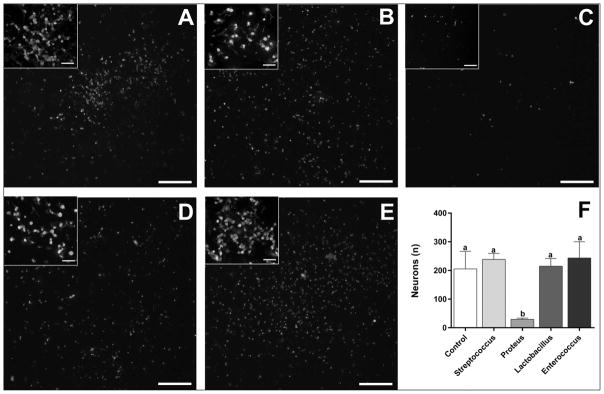
Obesity is associated with consumption of energy-dense diets and development of systemic inflammation. Gut microbiota play a role in energy harvest and inflammation and can influence the change from lean to obese phenotypes. The nucleus of the solitary tract (NTS) is a brain target for gastrointestinal signals modulating satiety and alterations in gut-brain vagal pathway may promote overeating and obesity. Therefore, we tested the hypothesis that high-fat diet‑induced changes in gut microbiota alter vagal gut-brain communication associated with increased body fat accumulation. Sprague-Dawley rats consumed a low energy‑dense rodent diet (LFD; 3.1 kcal/g) or high energy‑dense diet (HFD, 5.24 kcal/g). Minocycline was used to manipulate gut microbiota composition. 16S Sequencing was used to determine microbiota composition. Immunofluorescence against IB4 and Iba1 was used to determine NTS reorganization and microglia activation. Nodose ganglia from LFD rats were isolated and co-cultured with different bacteria strains to determine neurotoxicity. HFD altered gut microbiota with increases in Firmicutes/Bacteriodetes ratio and in pro-inflammatory Proteobacteria proliferation. HFD triggered reorganization of vagal afferents and microglia activation in the NTS, associated with weight gain. Minocycline-treated HFD rats exhibited microbiota profile comparable to LFD animals. Minocycline suppressed HFD‑induced reorganization of vagal afferents and microglia activation in the NTS, and reduced body fat accumulation. Proteobacteria isolated from cecum of HFD rats were toxic to vagal afferent neurons in culture. Our findings show that diet‑induced shift in gut microbiome may disrupt vagal gut‑brain communication resulting in microglia activation and increased body fat accumulation.


Figures

Fig. 1
Fig. 1
Minocycline treatment decreased energy intake and body fat accumulation induced by HFD. Seven days exposure to HFD (High fat diet) significantly increased energy intake (A, p<0.001; n=8/group) and body fat accumulation (B, p<0.05; n=8/group). After 21 days, when compared to saline-treated animals, HFD minocycline-treated rats exhibited a significant decrease in energy intake (A, p<0.05; n=4/group), body fat accumulation (B, p<0.05; n=4/group) and in body weight (C, p<0.05; n=4/group). Minocycline reduced the caloric intake in LFD rats only in the first week (A). However, between the 8th and 21st day of the experiment, this effect was abolished (A). Minocycline did not affect body fat accumulation and body weight of LFD rats (B, C). Bars represent the average value ±SEM; a, b, c – different letters denote significant differences.
Fig. 2
Fig. 2
Bacterial composition altered by HFD was improved by minocycline treatment. (A) Bacterial phyla abundance was quantified in fecal samples before dietary switch and treatment (baseline) and after 7 and 21 days on the different diets and treatments regimen. Firmicutes and Bacteriodetes were the most abundant bacterial phyla in all groups and at all time points. In all groups, there was a significant reduction in Verrucomicrobia abundance at day 7 (p<0.001) and day 21 (p<0.0001). Seven days of HFD were sufficient to induce a significant increase in Firmicutes (p<0.0001) and decrease in Bacteriodetes abundances (p<0.0001). Similar changes were observed after 21 days of HFD (A, Firmicutes, p<0.01; Bacteriodetes, p<0.001). Minocycline treatment improved HFD rats’ bacterial phyla profile with a significant increase in Bacteriodetes abundance after 7 days (A, p<0.05) and a significant reduction in Firmicutes (A, p<0.01) and increase in Bacteriodetes abundances (A, p<0.01) after 21 days. After 21 days of treatment, there were no significant differences in bacterial phyla abundance between the HFD /minocycline-treated rats and the LFD animals. Minocycline treatment did not significantly affect bacterial phyla abundance in LFD rats (Carvalho et al. 2012). a, b, c – different letters denote significant differences. (B) Negative and positive correlations were observed between energy intake and bacterial orders depleted (Bacteroidales) and enriched (Clostridiales) by HF feeding. Energy intake during the third week of experiment (14 to 21days) was correlated with order abundance measured at day 21 across diets and treatments. Bacteroidales abundance was negatively correlated with energy intake (r2=0.53, p<0.01) while Clostridiales abundance was positively correlated with intake (r2=0.47, p<0.01). (C and D) Bacterial orders that were significantly enriched or depleted by HFD and/or minocycline treatment (Log 2 fold changes from baseline). * – denotes significant difference from the LF_Saline group: * – p<0.05, ** – p<0.01, *** – p<0.001, **** – p<0.0001. (B) Consumption of HFD for 7 days led to significant increase in abundance of several bacterial orders belonging to the Firmicutes (Erysipelotrichales), Terenicutes (Entomoplasmatales, Mycoplasmatales), Proteobacteria (Rhodocyclales, Altermondales), Cyanobacteria (Nostocales, Chroococcales) and Verrucomicrobia (Puniceicoccales). HFD also led to significant depletion in Bacteriodetes orders Bacteroidales and Sphingobacteriales. C. Similar results were observed after 21 days with additional orders enriched (Enterobacteriales and Methylophilales, Proteobacteria) and depleted (Thermobaculales, Chloroflexi). (B) Minocycline treatment normalized HFD-induced dysbiosis. Seven days of minocycline exposure were sufficient to significantly reduce the HFD-induced proliferation of bacterial orders mentioned above, leading to other normalization of abundance or significant depletion (Nostocales, Chroococcales). Minocycline also prevented HFD-induced depletion in Bacteroidales and Sphingobacteriales. In LFD animals, minocycline alone significantly reduced the abundance of obesity-associated Puniceicoccales, Chroococcales and Clostridiales. (C) Similar results were observed after 21 days of minocycline treatment; minocycline led to normalization or depletion of HFD-associated bacterial orders and restored HFD-depleted orders. Minocycline alone led to a significant reduction in Erysipelotrichales (Firmicutes) and several Proteobacteria and Cyanobacteria orders, such as Rhodocyclales and Chroococcales.
Fig. 3
Fig. 3
Minocycline normalized HFD rats’ ‘microbiota profile and reduced HFD-induced metabolic endotoxemia. (A and B) PCA at the order level after 7 or 21 days of diet/minocycline treatments A. PCA showed that 7 days of HFD were sufficient to induce dysbiosis with a distinct microbiota profile while minocycline treatment led in a third profile with more variability, based on the diet consumed (LFD vs. HFD). B. HFD-induced dysbiosis was confirmed after 21 days of diet. Minocycline treatment led to normalization of the microbiota profile in HFD rats, with HF_Mino animals clustering with LF_Saline animals. There was no significant effect of minocycline treatment on the LF animals PCA scores. C. Circulating LPS levels in plasma after 7 or 21 days of diet/minocycline treatments. Seven days of HFD a non-significant increase in circulating LPS which became significant after 21 days of HFD (p<0.01). Minocycline treatment reduced circulating LPS in HFD rats and this effect was significant after 21 days (p<0.001). In LFD rats, minocycline treatment did not result in significant changes of LPS plasma levels at any time point. Bars represent the average value ±SEM; a, b – different letters denote significant differences.
Fig. 4
Fig. 4
High-fat diet-induced activation of microglia in the NTS and DMV was suppressed by minocycline treatment. (B–I) Representative images of Iba1 immunoreactivity in the hindbrain between bregma −13.10 and −14.10 mm. Immunostaining against Iba-1 revealed that HFD increased microglia activation in the NTS and the DMV. After 7 days on HFD microglia activation was observed only in the DMV (A, B–E) (p<0.05). After 21 days on HFD microglia activation was observed in both the DMV (p<0.01) and the NTS (p<0.01) (A, F–I). Minocycline treatment attenuated the HFD-induced increases in the microglia activation in both the DMV (p<0.001) and the NTS (p<0.01) at day 21 (A, F–I). Minocycline treatment did not significantly change the microglia activation in the DMV and the NTS in rats fed LFD at any studied time point. Bars represent the average value ±SEM; a, b – different letters denote significant differences. NTS: nucleus of the solitary tract; DMV: dorsal motor nucleus of the vagus; AP: area postrema; scale bar =200 μm.
Fig. 5
Fig. 5
HFD-induced vagal remodeling in the NTS and DMV was suppressed by minocycline. Binary analysis of the area fraction of IB4-labeled vagal afferents in the intermediate NTS and DMV revealed significant differences in the density of labeled afferent terminals between rats fed different diets. After 7 days HFD decreased the total IB4 labeling in the NTS (p<0.001) and the DMV (p<0.05) with compare to LFD (A, B–E). This effect was dampened by minocycline treatment only in the NTS but failed to reach significance. At day 21, we observed significant increase in density of IB4-labeled vagal afferents projecting to the NTS (p<0.001) and DMV (p<0.01) in HFD rats with compare to LFD rats (A, F–I). This effect was significantly decreased in both the NTS (p<0.001) and DMV (p<0.01) by minocycline treatment (A, F–I). There were no significant differences between LF_Saline and LF_Mino rats at any studied time point. Bars represent the average value ±SEM; a, b – different letters denote significant differences. NTS: nucleus of the solitary tract; DMV: dorsal motor nucleus of the vagus; AP: area postrema; scale bar =200 μm.
Fig. 6
Fig. 6
Gram-negative bacteria isolated from HFD rats significantly reduced the number of NG neurons in culture. Co-culture of NG primary sensory neurons isolated from LFD rats with Streptococcus mitis, Lactobacillus animalis or Enterococcus faecalis (B, D and E respectively; Firmicutes, Bacilli, Lactobacillales) isolated from HFD rats did not change the number of surviving neurons with compare to NG cultures without the bacteria (A, F). Adding Proteus mirablis (C; Proteobacteria, Gammaproteobacteria, Enterobacteriales), isolated from HFD rats, to NG cultures from LFD rats induced a dramatic loss of primary sensory neurons with compare to NG cultures without the bacteria (A, F, p<0.05). Inserts show high magnification images from the same section demonstrating neuronal morphology. Scale bar =200 μm or 20 μm in inserts. Bars represent the average value ±SEM; a, b – different letters denote significant differences.

Similar articles

Cited by

References

    1. Amin B, Hajhashemi V, Hosseinzadeh H. Minocycline potentiates the anti-hyperalgesic effect of ceftriaxone in CCI-induced neuropathic pain in rats. Res Pharm Sci. 2015;10:34–42. - PMC - PubMed
    1. Ballsmider LA, Vaughn AC, David M, Hajnal A, Di Lorenzo PM, Czaja K. Sleeve gastrectomy and Roux-en-Y gastric bypass alter the gut-brain communication. Neural Plast. 2015;2015:601985. - PMC - PubMed
    1. Benani A, Hryhorczuk C, Gouaze A, Fioramonti X, Brenachot X, Guissard C, Krezymon A, Duparc T, Colom A, Nedelec E, Rigault C, Lemoine A, Gascuel J, Gerardy-Schahn R, Valet P, Knauf C, Lorsignol A, Penicaud L. Food intake adaptation to dietary fat involves PSA-dependent rewiring of the arcuate melanocortin system in mice. J Neurosci. 2012;32:11970–11979. - PMC - PubMed
    1. Berthoud HR, Carlson NR, Powley TL. Topography of efferent vagal innervation of the rat gastrointestinal tract. Am J Physiol. 1991;260:R200–R207. - PubMed
    1. Berthoud HR, Shin AC, Zheng H. Obesity surgery and gut-brain communication. Physiol Behav. 2011;105:106–119. - PMC - PubMed

MeSH terms