. 2017 May 15;215(10):1558-1568.
doi: 10.1093/infdis/jix170.

Naturally Acquired Human Antibodies Against Reticulocyte-Binding Domains of Plasmodium vivax Proteins, PvRBP2c and PvRBP1a, Exhibit Binding-Inhibitory Activity

Affiliations

Naturally Acquired Human Antibodies Against Reticulocyte-Binding Domains of Plasmodium vivax Proteins, PvRBP2c and PvRBP1a, Exhibit Binding-Inhibitory Activity

Enna Dogra Gupta et al. J Infect Dis. .

Abstract

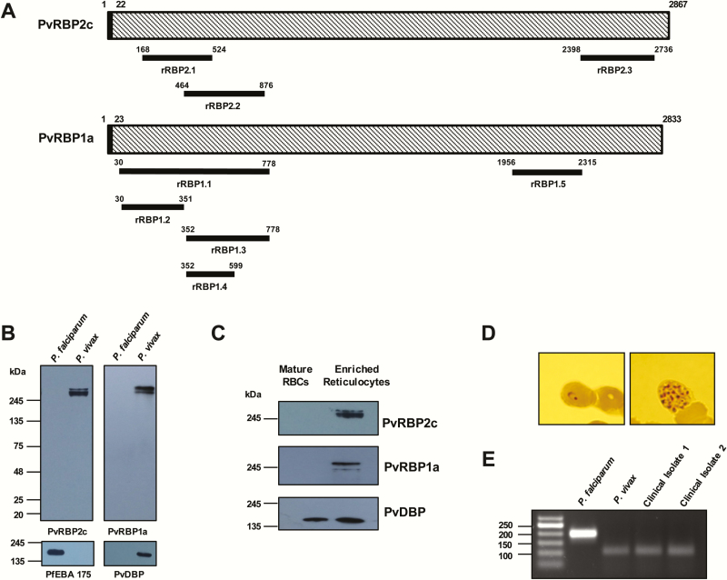
Background: Crucial gaps in our understanding of Plasmodium vivax reticulocyte invasion and protective immunity have hampered development of vivax vaccines. P. vivax exclusively invades reticulocytes that is mediated by the P. vivax reticulocyte-binding proteins (PvRBPs) specifically PvRBP2c and PvRBP1a. Vivax infections in Duffy-null individuals have suggested the evolution of alternate invasion pathways that may be mediated by the PvRBPs. Thus, PvRBPs appear as potential targets for efficacious P. vivax neutralization. However, there are limited data validating their vaccine efficacy. In the absence of vivax invasion assays, binding-inhibitory activity of antibodies has been reported to be associated with protection and a measure of vaccine potential.

Methods: -based analysis was performed of the PvRBP reticulocyte-binding properties and binding-inhibitory activity of specific anti-PvRBP2c/PvRBP1a human antibodies.

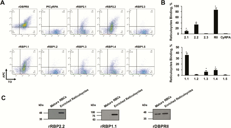
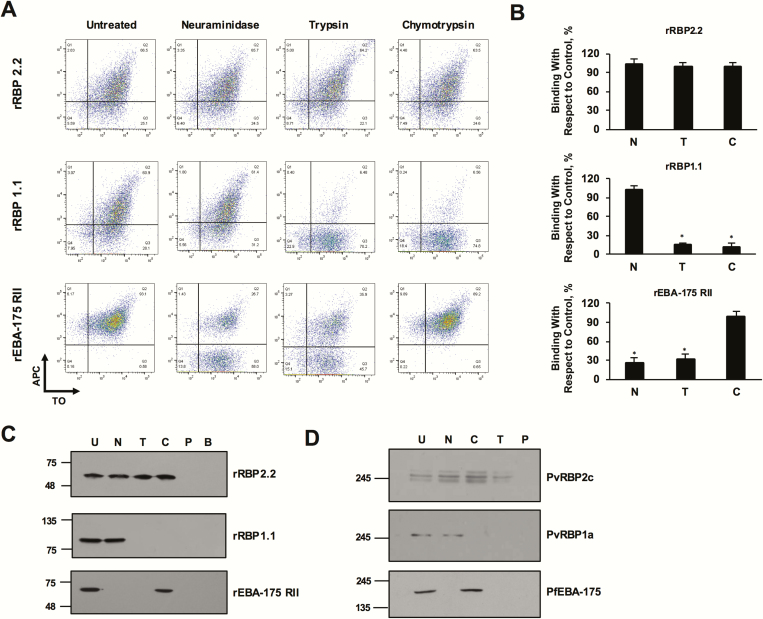
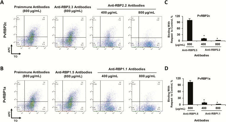
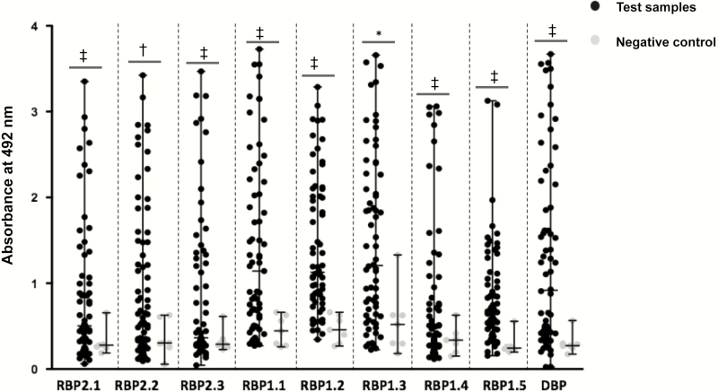
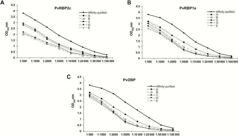
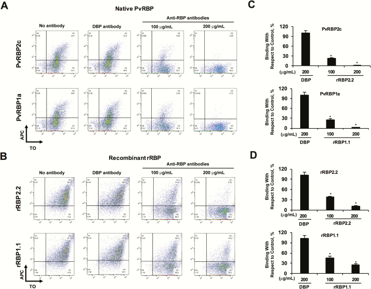
Results: PvRBP2c and PvRBP1a displayed a distinct reticulocyte-binding specificity, and their specific reticulocyte-binding domains were mapped within their N-terminal regions. Importantly, naturally acquired antibodies against the reticulocyte-binding domains efficaciously blocked reticulocyte binding of native PvRBPs, suggesting that the human immune system produced functional binding-inhibitory antibodies through exposure to vivax malaria.

Conclusions: Reticulocyte-binding domains of PvRBP2c/PvRBP1a are targets of naturally acquired binding-inhibitory antibodies, substantiating their promise as candidate antigens against which vaccine-inducible immunity could potentially be boosted through natural infections.

Keywords: binding-inhibitory antibodies; naturally acquired immunity; reticulocyte-binding proteins; vaccines.; vivax malaria.


Figures

Figure 1.
Figure 1.
Structure, expression, and reticulocyte-binding activity of the native Plasmodium vivax reticulocyte-binding proteins (PvRBPs). A, Schematic diagram of the PvRBP2c and PvRBP1a parasite proteins, highlighting the regions against which the recombinant proteins have been produced (black bars). B, Native PvRBP2c and PvRBP1a parasite proteins were detected by means of immunoblotting only in the P. vivax protein extracts by the specific anti-rRBP2.2 and anti-rRBP1.1 mice antibodies. Expression of native PfEBA-175 was detected only in the Plasmodium falciparum extract, confirming no cross-contamination of the P. vivax–infected samples with P. falciparum. Native P. vivax Duffy-binding protein (PvDBP) parasite protein was detected only in the P. vivax extract, confirming the specificity of the vivax sample. C, Binding of native PvRBP2c and PvRBP1a from the P. vivax culture supernatant was detected only with enriched reticulocytes and not with normal erythrocytes (red blood cells [RBCs]). Native PvDBP from the same culture supernatant bound with the same set of normal erythrocytes and enriched reticulocytes. Reticulocytes were enriched over a Percoll cushion. D, Blood film of the P. vivax–infected sample showing the P. vivax ring-stage and schizont-stage parasites. E, Identities of the P. vivax–infected samples were confirmed by means of nested polymerase chain reaction (PCR) amplification of 18S ribosomal RNA sequences, using primers specific for P. vivax and P. falciparum. The specific PCR-amplified DNA fragments from P. vivax and P. falciparum correspond to 100 and 200 base pairs (bp), respectively. Two representative P. vivax–infected clinical isolates are shown that correspond to the 100-bp size, confirming their identity. Abbreviations: PfEBA-175, P. falciparum erythrocyte binding antigen 175; rRBP, recombinant RBP.
Figure 2.
Figure 2.
Flow cytometry–based reticulocyte-binding activity of Plasmodium vivax reticulocyte-binding protein (PvRBP) 2c and PvRBP1a recombinant fragments. (A) Flow cytometry dot plots showing the binding of rRBP2.1, rRBP2.2, rRBP2.3, rDBPRII, and PfCyRPA (top) and rRBP1.1, rRBP1.2, rRBP1.3, rRBP1.4, rRBP1.5 (bottom) with enriched reticulocytes (thiazole orange [TO] positive). Reticulocyte binding was detected using the antibodies raised against the respective recombinant proteins followed by a secondary allophycocyanin (APC)–conjugated monoclonal antibody. Of the 3 PvRBP2c proteins, reticulocyte binding was observed for rRBP2.2 and rRBP2.1, with no binding observed for rRBP2.3. Of the 5 PvRBP1a protein constructs, reticulocyte binding was observed for rRBP1.1, rRBP1.3, and rRBP1.4, with no binding reported for rRBP1.2 and rRBP1.5. Binding of rDBPRII and PfCyRPA was analyzed as a positive and negative controls, respectively. B, Bar charts depicting the percentage reticulocyte binding of rRBP2.1, rRBP2.2, rRBP2.3, rDBPRII, PfCyRPA (top) and rRBP1.1, rRBP1.2, rRBP1.3, rRBP1.4, and rRBP1.5 (bottom). Error bars represent standard errors of the mean for 3 independent repeats. *P < .001. C, Binding of recombinant rRBP2.2 and rRBP1.1 was detected only with enriched reticulocytes and not with normal erythrocytes (red blood cells [RBCs]. rDBPRII bound with the same set of normal erythrocytes and enriched reticulocytes. Reticulocytes were enriched by magnetic sorting. Abbreviations: PfCyRPA, Plasmodium falciparum cysteine-rich protective antigen; rDBPRII, recombinant Duffy binding protein region II; rRBP, recombinant RBP.
Figure 3.
Figure 3.
Reticulocyte-binding specificity of rRBP2.2 and rRBP1.1 recombinant proteins. A, plots depicting rRBP2.2-binding reticulocytes in a neuraminidase-, chymotrypsin-, and trypsin-resistant manner. rRBP1.1 binding with reticulocytes (thiazole orange [TO] positive) was neuraminidase (N) resistant but sensitive to chymotrypsin (C) and trypsin (T). rEBA-175RII (recombinant receptor–binding domain of PfEBA-175) was analyzed as a control with the same set of enzymatically treated reticulocytes. rPfEBA-175RII bound untreated and chymotrypsin-treated cells but had reduced binding to the neuraminidase- and trypsin-treated cells. APC, allophycocyanin. B, Bar charts showing the percentage reticulocyte binding of rRBP2.2, rRBP21.1, and rEBA-175RII. Error bars represent standard errors of the mean for 3 independent repeats. *P < .001. C, Immunoblots depicting the binding of rRBP2.2, rRBP1.1, and rPfEBA-175RII to enzyme-treated reticulocytes. No protein was detected when cells were incubated with phosphate-buffered saline or when the magnetic beads were incubated with the proteins. B, CD71 magnetic beads control; U, untreated; C, chymotrypsin treated; N, neuraminidase treated; trypsin, P, phosphate-buffered saline control; T, trypsin treated. D, Immunoblots depicting the binding of native Plasmodium vivax reticulocyte-binding protein (PvRBP) 2c, PvRBP1a, and PfEBA-175 with the enzyme-treated reticulocytes. Reticulocytes were enriched by magnetic sorting. Abbreviations: PfEBA-175, Plasmodium falciparum erythrocyte binding antigen 175; rRBP, recombinant RBP.
Figure 4.
Figure 4.
Plasmodium vivax reticulocyte-binding protein (PvRBP) rat antibodies block reticulocyte binding of the native PvRBP parasite proteins. Flow cytometry dot plots depicting the inhibition of binding of native PvRBP2c (A) and PvRBP1a (B) in the presence of the respective anti-rRBP2.2/1.1 total rat immunoglobulin G (IgG; 400 and 800 µg/mL). APC, allophycocyanin; TO, thiazole orange. C, D, Bar charts showing the percentage of reticulocyte-binding inhibition of native PvRBP2c (C) and PvRBP1a (D) by anti-PvRBP IgG compared with preimmune rat IgG. The reticulocyte binding of the native PvRBP2c and PvRBP1a parasite proteins was potently blocked by the rRBP2.2 and rRBP1.1 rat antibodies (total IgG). As controls, reticulocyte binding of native PvRBP2c/PvRBP1a was analyzed in the presence of total rat IgG against the non-binders, rRBP2.3 and rRBP1.5. Anti-rat rRBP2.3/rRBP1.5 IgG failed to block the binding of native PvRBP2c and PvRBP1a proteins. Reticulocytes were enriched by magnetic sorting. *P < .001. Abbreviation: rRBP, recombinant RBP.
Figure 5.
Figure 5.
Detection of naturally acquired human antibody responses against Plasmodium vivax reticulocyte-binding protein (PvRBP) 2c, and PvRBP1a, and P. vivax Duffy-binding protein (PvDBP). Antibody responses were studied among 66 P. vivax–infected human plasma samples (dilution 1:200) against 9 recombinant proteins (8 PvRBP and 1 PvDBP). Seven healthy plasma samples from non–malaria-endemic regions with no history of malaria infections were used as controls. Bars indicate medians with ranges. *P < .05; †P < .01; ‡P < .001.
Figure 6.
Figure 6.
Analysis of the affinity-purified antibodies against Plasmodium vivax reticulocyte-binding protein (PvRBP) 2c (A), PvRBP1a (B), and P. vivax Duffy-binding protein (PvDBP) proteins (C). The enrichment of antigen-specific immunoglobulin Gs (IgGs) against rRBP2.2, rRBP1.1 and rDBPII were analyzed with enzyme-linked immunosorbent assay (ELISA). The ELISA results are plotted as optical density (OD) at 492 nm (y-axis) to compare concentrations of affinity-purified PvRBP2c (A), PvRBP1a (B), and PvDBP antibodies (C) and the 5 highest responders (plasma samples A–E). Antibody enrichment is reflected by the significantly higher OD values of the antigen-specific IgG in the affinity-purified samples compared with the highest responding individual plasma samples A-E. Abbreviations: DBP, Duffy binding protein; rRBP, recombinant RBP.
Figure 7.
Figure 7.
Purified Plasmodium vivax reticulocyte-binding protein (PvRBP)–specific human antibodies block reticulocyte binding of the native and recombinant PvRBPs. A, B, Flow cytometry dot plots depicting the inhibition of binding of native PvRBP parasite (A) proteins and rRBP2.2/rRBP1.1 (B) proteins in the presence of the respective anti-PvRBP2c/anti-PvRBP1a human immunoglobulin G (IgG; 100 and 200 µg/mL, respectively). APC, allophycocyanin; TO, thiazole orange. C, D, Bar charts showing the percentage inhibition of reticulocyte binding of native PvRBPs (C) and rRBPs (D) by the anti-PvRBP/P. vivax Duffy-binding protein (PvDBP) IgG compared with binding in the absence of IgG. The reticulocyte binding of the native PvRBP2c/PvRBP1a and recombinant rRBP2.2/rRBP1.1 were potently blocked by the respective PvRBP2c/PvRBP1a-specific human IgGs. As controls, reticulocyte binding of PvRBP2c/PvRBP1a was analyzed in the presence of PvDBP-specific purified human antibodies. Anti-PvDBP human IgG failed to block the reticulocyte binding of native PvRBP2c and PvRBP1a proteins. Reticulocytes were enriched by magnetic sorting. *P < .001. Abbreviation: rRBP, recombinant RBP.

References

    1. World Health Organization. World Malaria Report. 2015.
    1. Gething PW, Elyazar IR, Moyes CL, et al. A long neglected world malaria map: Plasmodium vivax endemicity in 2010. PLoS Negl Trop Dis 2012; 6:e1814. - PMC - PubMed
    1. Mueller I, Galinski MR, Baird JK, et al. Key gaps in the knowledge of Plasmodium vivax, a neglected human malaria parasite. Lancet Infect Dis 2009; 9:555–66. - PubMed
    1. Tanwar GS, Khatri PC, Sengar GS, et al. Clinical profiles of 13 children with Plasmodium vivax cerebral malaria. Ann Trop Paediatr 2011; 31:351–6. - PubMed
    1. Mueller I, Galinski MR, Tsuboi T, Arevalo-Herrera M, Collins WE, King CL. Natural acquisition of immunity to Plasmodium vivax: epidemiological observations and potential targets. Adv Parasitol 2013; 81:77–131. - PubMed

Publication types

MeSH terms