

The role of angiogenesis in Group 3 medulloblastoma pathogenesis and survival
- PMID: 28379574
- PMCID: PMC5570262
- DOI: 10.1093/neuonc/nox033
The role of angiogenesis in Group 3 medulloblastoma pathogenesis and survival
Abstract
Background: Of the 4 medulloblastoma subgroups, Group 3 is the most aggressive but the importance of angiogenesis is unknown. This study sought to determine the role of angiogenesis and identify clinically relevant biomarkers of tumor vascularity and survival in Group 3 medulloblastoma.
Methods: VEGFA mRNA expression and survival from several patient cohorts were analyzed. Group 3 xenografts were implanted intracranially in nude rats. Dynamic susceptibility weighted (DSC) MRI and susceptibility weighted imaging (SWI) were obtained. DSC MRI was used to calculate relative cerebral blood volume (rCBV) and flow (rCBF). Tumor vessel density and rat vascular endothelial growth factor alpha (VEGFA) expression were determined.
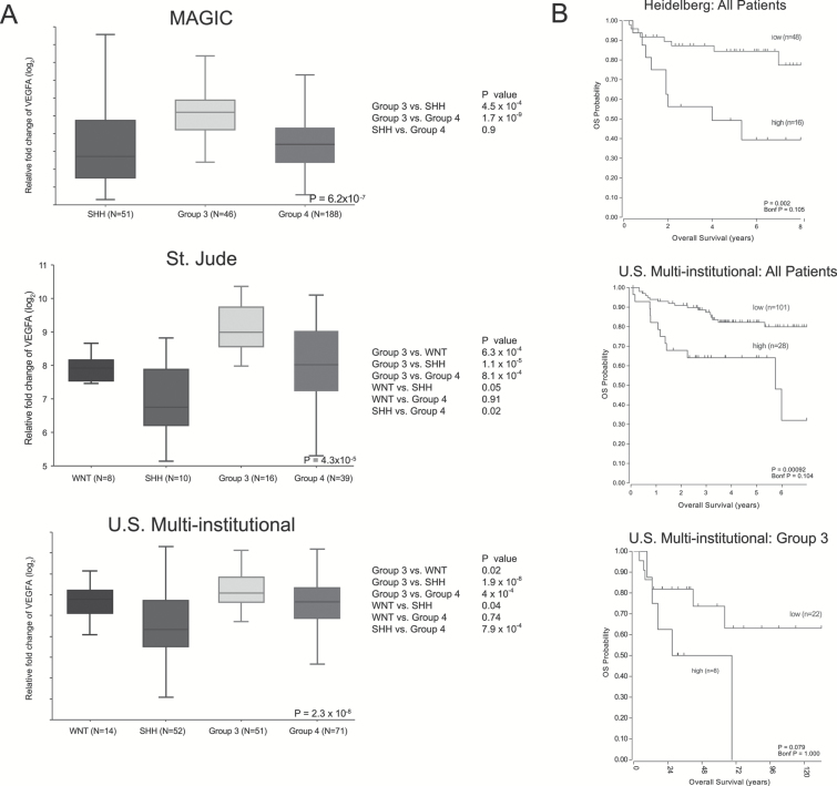
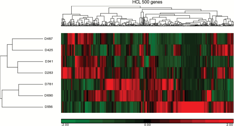
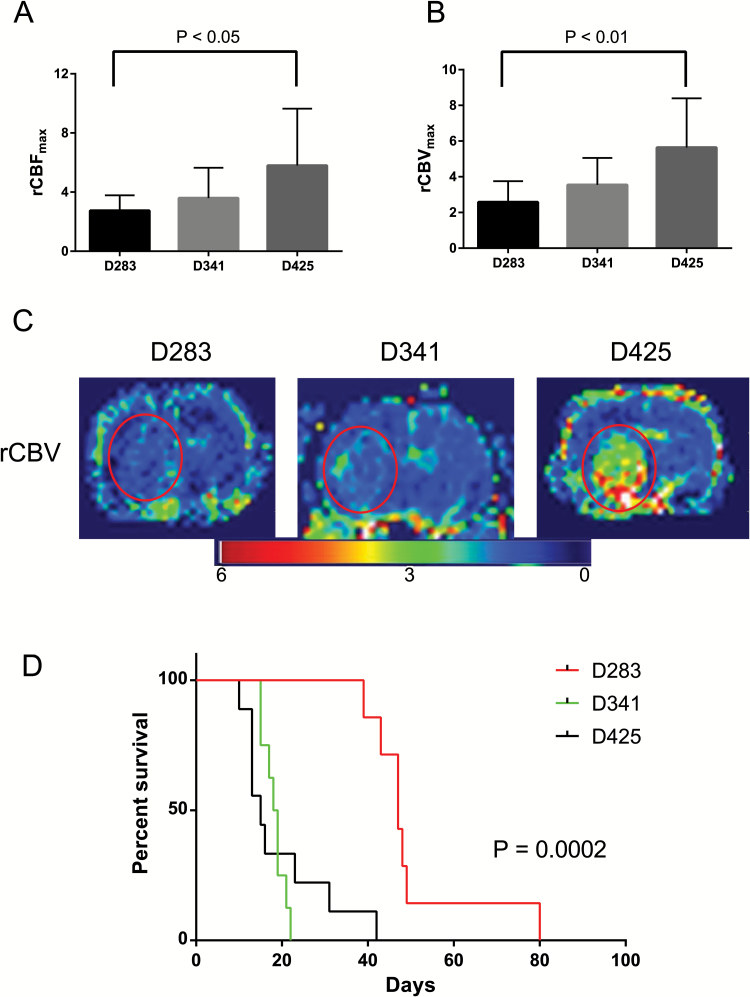
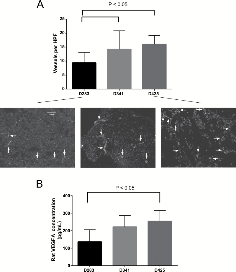
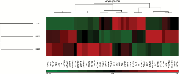
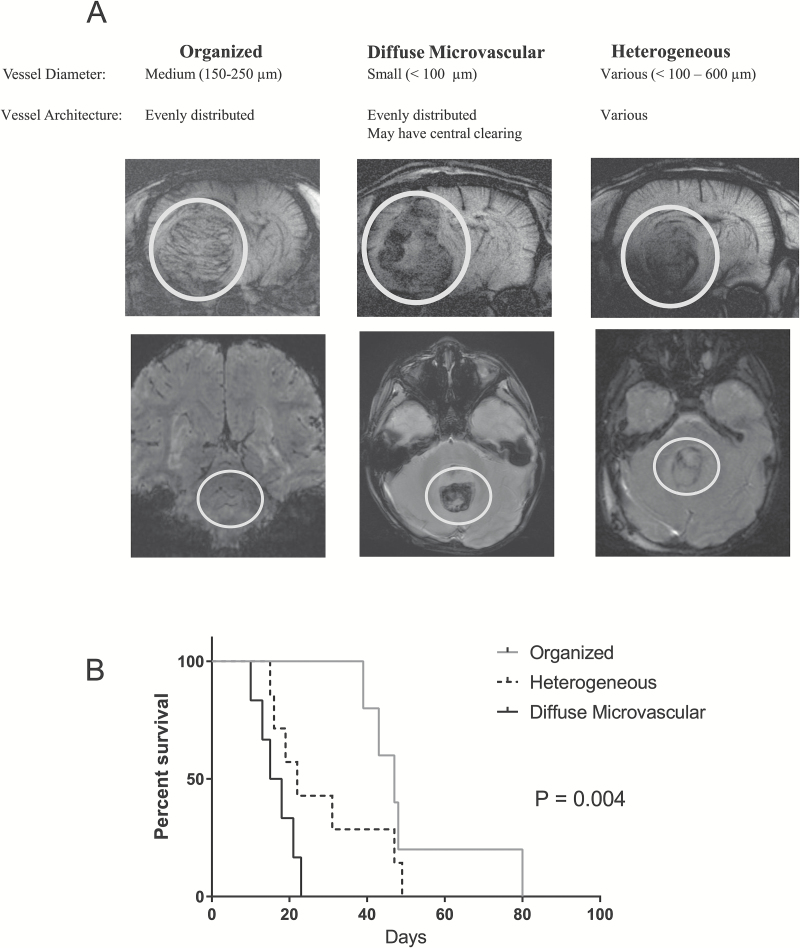
Results: Patient VEGFA mRNA levels were significantly elevated in Group 3 compared with the other subgroups (P < 0.001) and associated with survival. Xenografts D283, D341, and D425 were identified as Group 3 by RNA hierarchical clustering and MYC amplification. The D283 group had the lowest rCBV and rCBF, followed by D341 and D425 (P < 0.05). These values corresponded to histological vessel density (P < 0.05), rat VEGFA expression (P < 0.05), and survival (P = 0.002). Gene set enrichment analysis identified 5 putative genes with expression profiles corresponding with these findings: RNH1, SCG2, VEGFA, AGGF1, and PROK2. SWI identified 3 xenograft-independent categories of intratumoral vascular architecture with distinct survival (P = 0.004): organized, diffuse microvascular, and heterogeneous.
Conclusions: Angiogenesis plays an important role in Group 3 medulloblastoma pathogenesis and survival. DSC MRI and SWI are clinically relevant biomarkers for tumor vascularity and overall survival and can be used to direct the use of antivascular therapies for patients with Group 3 medulloblastoma.
Keywords: Group 3; VEGF; angiogenesis; dynamic susceptibility contrast MRI; medulloblastoma.
© The Author(s) 2017. Published by Oxford University Press on behalf of the Society for Neuro-Oncology. All rights reserved. For permissions, please e-mail: journals.permissions@oup.com
Figures
Similar articles
-
Differences in vascular endothelial growth factor receptor expression and correlation with the degree of enhancement in medulloblastoma.J Neurosurg Pediatr. 2014 Aug;14(2):121-8. doi: 10.3171/2014.4.PEDS13244. Epub 2014 Jun 6. J Neurosurg Pediatr. 2014. PMID: 24905841
-
Dynamic susceptibility contrast-enhanced MRI with USPIO in evaluating angiogenesis of the peri-infarction zones in subacute ischemic stroke in a permanent middle cerebral artery occlusion rat model.Acta Radiol. 2024 Dec;65(12):1529-1539. doi: 10.1177/02841851241290646. Epub 2024 Oct 24. Acta Radiol. 2024. PMID: 39449316
-
Valproic Acid prolongs survival time of severe combined immunodeficient mice bearing intracerebellar orthotopic medulloblastoma xenografts.Clin Cancer Res. 2006 Aug 1;12(15):4687-94. doi: 10.1158/1078-0432.CCR-05-2849. Clin Cancer Res. 2006. PMID: 16899619
-
Childhood Medulloblastoma Revisited.Top Magn Reson Imaging. 2018 Dec;27(6):479-502. doi: 10.1097/RMR.0000000000000184. Top Magn Reson Imaging. 2018. PMID: 30516696 Review.
-
Medulloblastoma with Atypical Dynamic Imaging Changes: Case Report with Literature Review.World Neurosurg. 2017 Sep;105:1036.e1-1036.e3. doi: 10.1016/j.wneu.2017.06.058. Epub 2017 Jun 16. World Neurosurg. 2017. PMID: 28625899 Review.
Cited by
-
The Current Landscape of Targeted Clinical Trials in Non-WNT/Non-SHH Medulloblastoma.Cancers (Basel). 2022 Jan 28;14(3):679. doi: 10.3390/cancers14030679. Cancers (Basel). 2022. PMID: 35158947 Free PMC article. Review.
-
Targeting Angiogenic Factors for the Treatment of Medulloblastoma.Curr Treat Options Oncol. 2022 Jun;23(6):864-886. doi: 10.1007/s11864-022-00981-1. Epub 2022 Apr 12. Curr Treat Options Oncol. 2022. PMID: 35412196 Review.
-
Addressing blood-brain-tumor-barrier heterogeneity in pediatric brain tumors with innovative preclinical models.Front Oncol. 2023 Jan 26;13:1101522. doi: 10.3389/fonc.2023.1101522. eCollection 2023. Front Oncol. 2023. PMID: 36776301 Free PMC article. Review.
-
Potential therapeutic target secretogranin II might cooperate with hypoxia-inducible factor 1α in sunitinib-resistant renal cell carcinoma.Cancer Sci. 2023 Oct;114(10):3946-3956. doi: 10.1111/cas.15914. Epub 2023 Aug 6. Cancer Sci. 2023. PMID: 37545017 Free PMC article.
-
[High expression of secretogranin II increases oxaliplatin resistance of colorectal cancer cells].Nan Fang Yi Ke Da Xue Xue Bao. 2023 Oct 20;43(10):1657-1664. doi: 10.12122/j.issn.1673-4254.2023.10.02. Nan Fang Yi Ke Da Xue Xue Bao. 2023. PMID: 37933640 Free PMC article. Chinese.
References
-
- Huber H, Eggert A, Janss AJ, et al. Angiogenic profile of childhood primitive neuroectodermal brain tumours/medulloblastomas. Eur J Cancer. 2001;37(16):2064–2072. - PubMed
-
- Jain RK, di Tomaso E, Duda DG, Loeffler JS, Sorensen AG, Batchelor TT. Angiogenesis in brain tumours. Nat Rev Neurosci. 2007;8(8):610–622. - PubMed
-
- Li VW, Folkerth RD, Watanabe H, et al. Microvessel count and cerebrospinal fluid basic fibroblast growth factor in children with brain tumours. Lancet. 1994;344(8915):82–86. - PubMed
-
- Nagase T, Nagase M, Machida M, Fujita T. Hedgehog signalling in vascular development. Angiogenesis. 2008;11(1):71–77. - PubMed
MeSH terms
Substances
Grants and funding
LinkOut - more resources
Full Text Sources
Other Literature Sources
Miscellaneous
