. 2017 Apr 4;25(4):823-837.e8.
doi: 10.1016/j.cmet.2017.03.006.

A Class of Reactive Acyl-CoA Species Reveals the Non-enzymatic Origins of Protein Acylation

Affiliations

A Class of Reactive Acyl-CoA Species Reveals the Non-enzymatic Origins of Protein Acylation

Gregory R Wagner et al. Cell Metab. .

Abstract

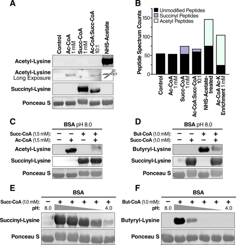
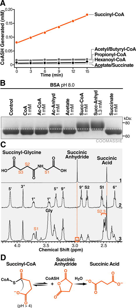
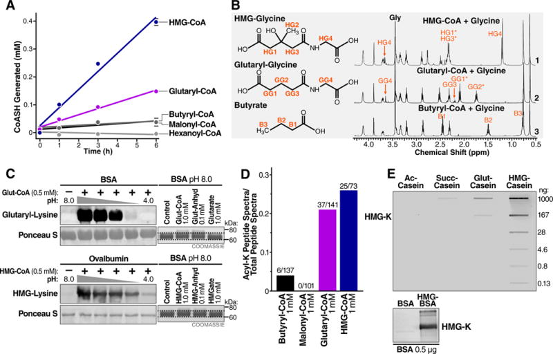
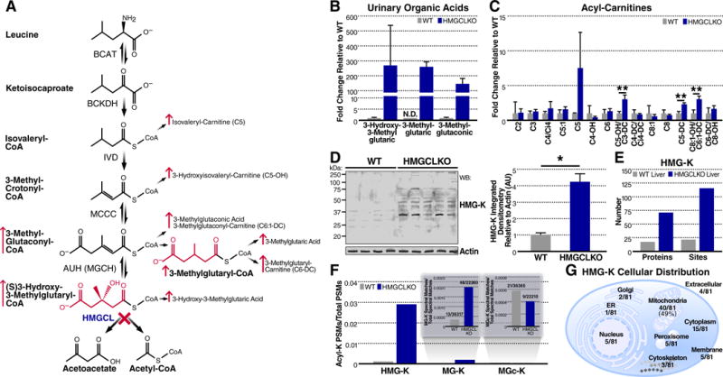
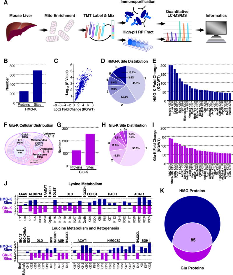
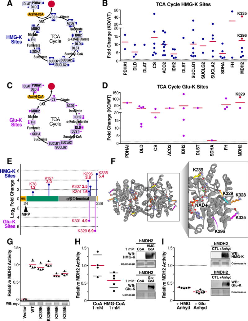
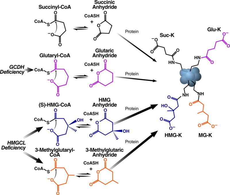
The mechanisms underlying the formation of acyl protein modifications remain poorly understood. By investigating the reactivity of endogenous acyl-CoA metabolites, we found a class of acyl-CoAs that undergo intramolecular catalysis to form reactive intermediates that non-enzymatically modify proteins. Based on this mechanism, we predicted, validated, and characterized a protein modification: 3-hydroxy-3-methylglutaryl(HMG)-lysine. In a model of altered HMG-CoA metabolism, we found evidence of two additional protein modifications: 3-methylglutaconyl(MGc)-lysine and 3-methylglutaryl(MG)-lysine. Using quantitative proteomics, we compared the "acylomes" of two reactive acyl-CoA species, namely HMG-CoA and glutaryl-CoA, which are generated in different pathways. We found proteins that are uniquely modified by each reactive metabolite, as well as common proteins and pathways. We identified the tricarboxylic acid cycle as a pathway commonly regulated by acylation and validated malate dehydrogenase as a key target. These data uncover a fundamental relationship between reactive acyl-CoA species and proteins and define a new regulatory paradigm in metabolism.

Keywords: acyl-CoA; chemical biology; non-enzymatic; post-translational modifications; protein acylation; sirtuins.


Conflict of interest statement

COMPETING FINANCIAL INTERESTS

The authors declare no competing financial interests.

Figures

Figure 1
Figure 1
The primary citric acid cycle intermediate succinyl-CoA is an efficient acylating agent. (A) Western blot for acetyl-lysine or succinyl-lysine residues on BSA acylated in vitro with acetyl-CoA and succinyl-CoA. BSA was incubated with different concentrations of acyl-CoAs, NHS-acetate, or immunoenriched with an acetyl-lysine antibody. The acetyl-lysine long exposure was performed after the NHS-acetate treated sample lane was removed. (B) Total peptide spectral matches (PSMs) of unmodified (non-acylated) or acylated (acetyl or succinyl) peptides identified from samples in (A) using liquid chromatography tandem mass spectrometry. (C) Protein acylation by succinyl-CoA outcompetes acylation by acetyl-CoA at equimolar concentration. (D) Protein acylation by succinyl-CoA outcompetes acylation by butyryl-CoA at equimolar concentrations. (E) Western blot for succinyl-lysine or (F) butyryl-lysine residues on BSA acylated by succinyl-CoA or butyryl-CoA over pH range 8–4. The middle lanes are pH 7,6, and 5 from left to right. The nitrocellulose membranes used for immunoblotting were stained with the non-specific protein marker Ponceau S. WB, Western blot.
Figure 2
Figure 2
Succinyl-CoA undergoes intramolecular catalysis-mediated formation of a highly reactive cyclic anhydride intermediate. (A) Equimolar acyl-CoAs were monitored for the formation of free CoASH with Ellman’s reagent. (B) Comparison of mass shifts induced by incubation of BSA with acetyl-CoA, acetic anhydride, succinyl-CoA, or succinic anhydride. (C) Expansions of proton NMR spectra demonstrating the formation of succinyl-glycine from succinyl-CoA and glycine at pH 5.0. (1) Succinic anhydride and succinic acid at pH 5.0. (2) Succinyl-CoA with assignments based on (Laurieri et al., 2014). The S1 and S2 peaks correspond to the succinyl methylene groups. (3) Succinyl-CoA with glycine at pH 5.0. The reaction produces succinyl-glycine with assignments indicated. Succinyl-CoA generates a small peak at 2.96 ppm that is consistent with the transient formation of the cyclic succinic anhydride intermediate. (D) Model of succinyl-CoA undergoing intramolecular catalysis to form a reactive anhydride intermediate.
Figure 3
Figure 3
Endogenous five-carbon dicarboxyl acyl-CoAs undergo intramolecular catalysis and anhydride formation. (A) Equimolar acyl-CoAs were monitored for the formation of free CoASH with Ellman’s reagent. (B) Proton NMR spectra of acyl-glycine formation after incubating (1) HMG-, (2) glutaryl-, and (3) butyryl-CoA with glycine at pH 7.0. (C) (Left) the acylation of BSA by glutaryl-CoA and HMG-CoA over pH range 8–4. The middle lanes are pH 7,6, and 5 from left to right. The nitrocellulose membranes used for immunoblotting were stained with Ponceau S for loading. (Right) Comparison of mass shifts induced by incubation of BSA with glutaryl-CoA, HMG-CoA and their related anhydrides and acids. (D) BSA was acylated in vitro with butyryl-CoA, malonyl-CoA, glutaryl-CoA, or HMG-CoA and subjected to liquid chromatography tandem mass spectrometry. Fraction of acylated peptide spectra (butyryl, malonyl, glutaryl, and hydroxymethylglutaryl) identified out of the total number of peptide spectra (acyl-lysine and non-acyl-lysine) is shown. (E) (Top) Sensitivity and specificity of purified HMG-K antiserum was tested against the indicated amounts of casein after acetylation (ac-casein), succinylation (suc-casein), glutarylation (glut-casein), and HMGylation (HMG-casein). (Bottom) The hydroxymethylglutaryl-lysine (HMG-K) antiserum recognizes BSA chemically modified to contain hydroxymethylglutaryl-lysine modifications, but not unmodified BSA.
Figure 4
Figure 4
Lysine HMGylation is an endogenous protein modification. (A) Schematic of leucine catabolism indicating the step compromised in HMGCL deficiency and consequent elevation of acyl-CoAs, organic acids, and acyl-carnitines. (B) Relative quantification of select organic acids derived from leucine catabolism from urine in WT and HMGCLKO mice. Levels of 3-methylglutaric acid in WT urine were not detectable (N.D.), therefore, a relative comparison is depicted with the WT value set to 0.1 and fold-change derived accordingly. Data represent mean ± SEM (C) Relative quantification of selected acyl-carnitines in WT and HMGCLKO mouse livers. Multiple t-tests were used to evaluate statistical significance without correction for multiple comparisons. Data represent mean ± SD **P ≤ 0.01. (D) Western blot probing for HMG-lysine modifications and actin in WT (n=4) and HMGCLKO (n=5) liver lysates. At right, quantification of integrated densitometry in the Western blot normalized to actin. A two-tailed Mann-Whitney U test was used to evaluate statistical significance. Data represent mean ± SD *P ≤ 0.05. (E) Summary of HMG-K containing peptides and proteins identified in WT and HMGCLKO livers (F) The fraction of peptide spectral matches identified in the HMG-K immunoprecipitation containing HMG-lysine, 3-methyglutaryl-lysine (MG-K), and 3-methylglutaconyl-lysine (MGc-K) modifications. (G) The cellular distribution of HMG-K containing proteins identified in the HMG-K immunoprecipitation.
Figure 5
Figure 5
Quantitative proteomics reveals the breadth and depth of the mitochondrial HMGylome. (A) Proteomic workflow. (B) Summary of HMGylated proteins and sites identified through quantitative proteomics (C) Volcano plot of log2 fold-change in HMGylation at sites identified in the HMGCLKO mitochondria relative to WT mitochondria and their associated −log10 P values. (D) Distribution of the number of HMGylation sites on HMGylated proteins. (E) Protein lysine residues displaying the greatest increases in HMGylation in HMGCLKO mitochondria. (F) Cellular distribution of glu-K proteins identified in GCDHKO mouse liver. (G) Summary of glutarylated proteins and sites identified through quantitative proteomics. (H) Distribution of the number of glutarylation sites on glutarylated proteins. (I) Protein lysine residues displaying the greatest increases in glutarylation in GCDHKO liver, relative to WT. (J) Fold-change in HMGylation (blue) and glutarylation (purple) at specific sites on proteins of lysine metabolism and leucine metabolism and ketogenesis in HMGCLKO and GCDHKO samples, relative to their respective WT controls. (K) The number of proteins containing a detected site of lysine HMGylation, lysine glutarylation, or both.
Figure 6
Figure 6
Acylation reduces MDH2 activity. (A) Summary of TCA Cycle proteins with elevated HMGylation in HMGCLKO liver mitochondria as determined through quantitative TMT proteomics. Each ball-stick represents one unique quantifiable site of HMGylation. (B) Site-specific relative fold-change in HMGylation occurring on proteins in the TCA cycle in HMGCLKO mitochondria relative to WT. HMGylation sites found on MDH2 that were mutated are colored in red. Horizontal lines represent the average fold change for all sites within a protein. (C) Similar to (A), but showing sites of glutarylation found GCDHKO mouse liver (D) Similar to (B), but showing relative fold change in glutarylation sites found in GCDHKO mouse liver. (E) The protein domains plotted in the amino acid sequence of MDH2 showing the quantified sites of HMGylation and glutarylation as blue and purple ball-stick diagrams, respectively. The height of the mark represents the relative fold increase in modification at that site. (F) Structural analysis of human MDH2 crystal structure (PDB 2DFD) modeling lysines 239, 296, 328/9 and 335 and relevant intramolecular interactions. The dashed green lines indicate hydrogen bonds and the dashed orange lines indicate electrostatic interactions. (G) Relative activity of recombinant human wild-type MDH2 and mutant MDH2 containing lysine (K) to glutamate (E) mutations overexpressed in 293T cells and activity was measured in cell lysates; values corrected for vector control activity and MDH2 protein abundance. Below, a representative Western blot probing for myc in cell lysate containing the overexpressed MDH2-myc constructs. (H) Relative catalytic activity of human recombinant MDH2 after incubation with coenzyme A (CoA) (n = 4) or 3-hydroxy-3-methylglutaryl-CoA (HMG-CoA) (n = 5) for 12 hours. At right, Western blots for HMG-K or glu-K modifications in CoA, HMG-CoA, and glutaryl-CoA treated hMDH2 samples. (I) Catalytic activity of human recombinant malate dehydrogenase 2 (hMDH2) after incubation with hydroxymethylglutaric anhydride or glutaric anhydride for 5 minutes (n = 5 for both conditions) relative to untreated MDH2. At right, Western blots for HMG-K or glu-K modifications in HMG anhydride and glutaric anhydride treated hMDH2 samples. Data represent mean ± standard deviation. WB, Western blot.
Figure 7
Figure 7
Protein acylation by four- and five-carbon dicarboxylic acyl-CoAs. The terminal carboxylic acids in succinyl-CoA, glutaryl-CoA, HMG-CoA, and 3-methylglutaryl-CoA facilitate non-enzymatic lysine acylation through intramolecular general base catalysis-mediated formation of a highly reactive cyclic anhydride intermediate. In a mouse model of the human disorder HMG-CoA lyase deficiency, HMG-CoA and 3-methylglutaryl-CoA accumulate and, consequently, protein lysine HMGylation and 3-MGylation are elevated. In a mouse model of the human disorder glutaric academia (GA I), glutaryl-CoA accumulates and, consequently, protein lysine glutarylation is elevated.

Comment in

  • Cycling around Lysine Modifications.
    Kumar S, Lombard DB. Kumar S, et al. Trends Biochem Sci. 2017 Jul;42(7):501-503. doi: 10.1016/j.tibs.2017.05.006. Epub 2017 Jun 6. Trends Biochem Sci. 2017. PMID: 28595832 Free PMC article.

References

    1. Anderson KA, Green MF, Huynh FK, Wagner GR, Hirschey MD. SnapShot: Mammalian Sirtuins. Cell. 2014;159:956–956 e951. - PMC - PubMed
    1. Anderson KA, Huynh FK, Fisher-Wellman K, Stuart JD, Peterson BS, Douros JD, Wagner GR, Thompson JW, Madsen AS, Green MF, et al. SIRT4 Is a Lysine Deacylase that Controls Leucine Metabolism and Insulin Secretion. 2017 Submitted. - PMC - PubMed
    1. Bender ML. Mechanisms of Catalysis of Nucleophilic Reactions of Carboxylic Acid Derivatives. Chem Rev. 1960;60:53–113.
    1. Brooks BR, Brooks CL, 3rd, Mackerell AD, Jr, Nilsson L, Petrella RJ, Roux B, Won Y, Archontis G, Bartels C, Boresch S, et al. CHARMM: the biomolecular simulation program. J Comput Chem. 2009;30:1545–1614. - PMC - PubMed
    1. Buckel W. Acetic anhydride: an intermediate analogue in the acyl-exchange reaction of citramalate lyase. Eur J Biochem. 1976;64:263–267. - PubMed