Clinical Trial
. 2017 Apr 5;6(4):e003918.
doi: 10.1161/JAHA.116.003918.

Phase I Clinical Trial of Autologous Stem Cell-Sheet Transplantation Therapy for Treating Cardiomyopathy

Affiliations
Clinical Trial

Phase I Clinical Trial of Autologous Stem Cell-Sheet Transplantation Therapy for Treating Cardiomyopathy

Shigeru Miyagawa et al. J Am Heart Assoc. .

Abstract

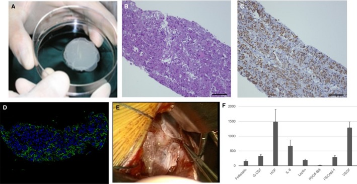
Background: When transplanted into failing heart, autologous somatic tissue-derived cells yield functional recovery via paracrine effects that enhance native regeneration. However, the therapeutic effects are modest. We developed a method in which scaffold-free cell sheets are attached to the epicardial surface to maximize paracrine effects. This Phase I clinical trial tested whether transplanting autologous cell-sheets derived from skeletal muscle is feasible, safe, and effective for treating severe congestive heart failure.

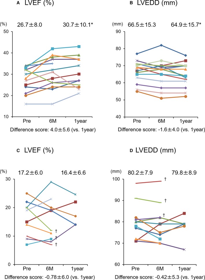
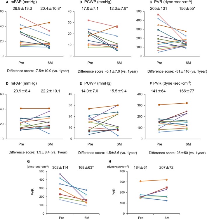
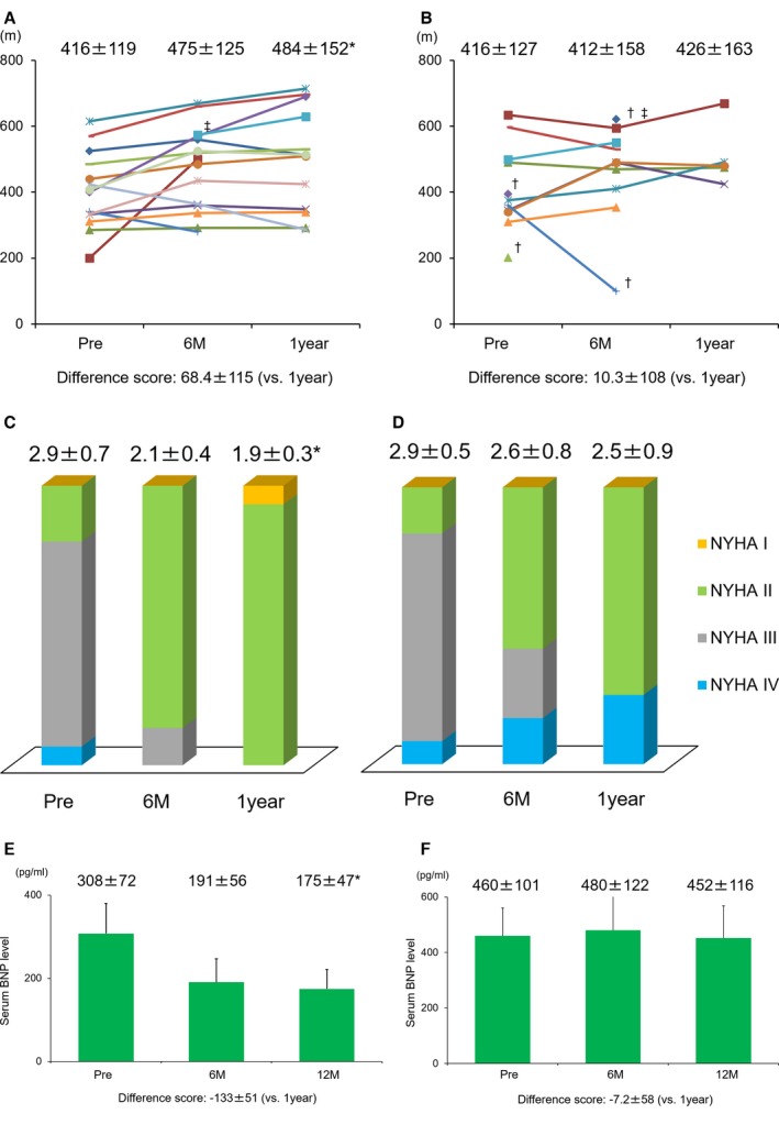
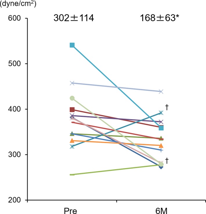
Methods and results: Fifteen ischemic cardiomyopathy patients and 12 patients with dilated cardiomyopathy, who were in New York Heart Association functional class II or III and had been treated with the maximum medical and/or interventional therapies available, were enrolled. Scaffold-free cell sheets of 3 to 9×10⁸ cells derived from autologous muscle were transplanted over the LV free wall via left thoracotomy, without additional interventional treatments. There were no procedure-related major complications during follow-up. The majority of the ischemic cardiomyopathy patients showed marked symptomatic improvement in New York Heart Association classification (pre: 2.9±0.5 versus 6 months: 2.1±0.4, P<0.01; 1 year: 1.9±0.3, P<0.01) and the Six-Minute Walk Test with significant reduction of serum brain natriuretic peptide level (pre: 308±72 pg/mL versus 6 months: 191±56 versus 1 year: 182±46, P<0.05), pulmonary artery pressure, pulmonary capillary wedge pressure, pulmonary vein resistance, and left ventricular wall stress after transplantation instead of limited efficacy in dilated cardiomyopathy patients.

Conclusions: Cell-sheet transplantation as a sole therapy was feasible for treating cardiomyopathy. Promising results in the safety and functional recovery warrant further clinical follow-up and larger studies to confirm this treatment's efficacy for severe congestive heart failure.

Clinical trial registration: URL: http://www.umin.ac.jp/english/. Unique identifier: UMIN000003273.

Keywords: autologous stem cell‐sheet; cellular transplantation; ejection fraction; growth factors and cytokines; ischemic cardiomyopathy; myocardial regeneration.


Figures

Figure 1
Figure 1
Characterization of implanted autologous skeletal stem‐cell sheets. A, The appearance of cell sheets. B, Hematoxylin and eosin staining. C, Desmin staining. D, Fibronectin staining. E, Cell sheets were implanted to LV free wall via left thoracotomy. F, In vitro study; various cytokines were detected in supernatant of cell sheet. G‐CSF indicates granulocyte colony‐stimulating factor; HGF, hepatocyte growth factor; IL‐8, interleukin‐8; PDGFBB, platelet‐derived growth factor‐BB; PECAM‐1, platelet endothelial cell adhesion molecule; VEGF, vascular endothelial growth factor.
Figure 2
Figure 2
Echocardiographic analysis of cardiac function. A and B, Change of LVEF and LVEDD in the patients with ischemic etiology. C and D, Change of LVEF and LVEDD in the patients with nonischemic etiology. Each color bar corresponds to an individual patient. *vs pre P<0.05 (Friedman's test). Examination was not performed because deterioration of cardiac function resulted in LVAD implantation or dependence on continuous catecholamine infusion within 1 year after treatment. LVAD indicates left ventricular assist device; LVEDD, left ventricular end‐diastolic dimension; LVEF, left ventricular ejection fraction.
Figure 3
Figure 3
Pressure study evaluated by Swan‐Ganz catheter. Each color bar corresponds to an individual patient. A through C, Pressure study in the patients with ischemic etiology. D through F, Pressure study in the patients with nonischemic etiology. G, Change of PVR in the patients with ischemic etiology and preoperative pulmonary hypertension. H, Change of PVR in the patients with nonischemic etiology and preoperative pulmonary hypertension. mPAP indicates mean pulmonary artery pressure; PCWP, pulmonary capillary wedge pressure; PVR, pulmonary vascular resistance. *vs pre P<0.05 (Wilcoxon signed‐rank test).
Figure 4
Figure 4
Exercise capacity and symptoms. A, The changes of exercise capacity evaluated by 6‐Minute Walk Test in the ischemic etiology. B, The changes of exercise capacity evaluated by 6‐Minute Walk Test in the nonischemic etiology. Each color bar in A and B corresponds to an individual patient. C, The changes of symptoms evaluated by NYHA functional classification in the ischemic etiology. D, The changes of symptoms evaluated by NYHA functional classification in the nonischemic etiology. E, The time course changes in serum level of BNP in the ischemic etiology (mean±SE). F, The time course changes in serum level of BNP in the nonischemic etiology (mean±SE). *vs pre P<0.05 (Friedman's test). No examination was performed, because of deterioration of cardiac function after the treatment. No examination was performed preoperatively because of depending on continuous catecholamine infusion. BNP indicates brain natriuretic peptide; NYHA, New York Heart Association.
Figure 5
Figure 5
End‐systolic wall stress evaluated by MDCT. Each color bar corresponds to an individual patient. The changes of end‐systolic wall stress in the ischemic etiology: MDCT evaluation was not performed in 2 patients because of renal dysfunction. Increasing of ESS was observed only in 2 patients in the ischemic etiology. *vs pre P<0.05 (Wilcoxon signed‐rank test). ESS indicates end‐systolic wall stress; MDCT, cine‐multidetector computed tomography.
Figure 6
Figure 6
Clinical outcome of late periods. A, Freedom from all‐cause death in both etiologies. B, Freedom from cardiac death in both etiologies. C, Freedom from heart failure event requiring in‐hospital treatment. GI bleeding indicates gastrointestinal bleeding; HF event, heart failure event requiring in‐hospital treatment.

References

    1. Miyagawa S, Roth M, Saito A, Sawa Y, Kostin S. Tissue‐engineered cardiac constructs for cardiac repair. Ann Thorac Surg. 2011;91:320–329. - PubMed
    1. Miyagawa S, Saito A, Sakaguchi T, Yoshikawa Y, Yamauchi T, Imanishi Y. Impaired myocardium regeneration with skeletal cell sheets–a preclinical trial for tissue‐engineered regeneration therapy. Transplantation. 2010;90:364–372. - PubMed
    1. Memon IA, Sawa Y, Fukushima N, Matsumiya G, Miyagawa S, Taketani S, Sakakida S, Kondoh H, Aleshin AN, Shimizu T, Okano T, Matsuda H. Repair of impaired myocardium by means of implantation of engineered autologous myoblast sheets. J Thorac Cardiovasc Surg. 2005;130:1333–1341. - PubMed
    1. Menasché P, Vanneaux V, Hagège A, Bel A, Cholley B, Cacciapuoti I, Parouchev A, Benhamouda N, Tachdjian G, Tosca L, Trouvin JH, Fabreguettes JR, Bellamy V, Guillemain R, Suberbielle Boissel C, Tartour E, Desnos M, Larghero J. Human embryonic stem cell‐derived cardiac progenitors for severe heart failure treatment: first clinical case report. Eur Heart J. 2015;36:2011–2017. - PubMed
    1. Narita T, Shintani Y, Ikebe C, Kaneko M, Campbell NG, Coppen SR, Uppal R, Sawa Y, Yashiro K, Suzuki K. The use of scaffold‐free cell sheet technique to refine mesenchymal stromal cell‐based therapy for heart failure. Mol Ther. 2013;21:860–867. - PMC - PubMed

Publication types