. 2017 Jul 1;118(1):254-266.
doi: 10.1152/jn.00825.2016. Epub 2017 Apr 5.

Parallel processing of internal and external feedback in the spinocerebellar system of primates

Affiliations

Parallel processing of internal and external feedback in the spinocerebellar system of primates

Oren Cohen et al. J Neurophysiol. .

Abstract

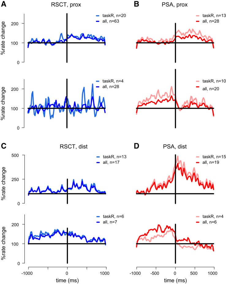
Cerebellar control of voluntary movements is achieved by the integration of external and internal feedback information to adjust and correct properly ongoing actions. In the forelimb of primates, rostral-spinocerebellar tract (RSCT) neurons are thought to integrate segmental, descending, and afferent sources and relay upstream a compound signal that contains both an efference copy of the spinal-level motor command and the state of the periphery. We tested this hypothesis by implanting stimulating electrodes in the superior cerebellar peduncle and recording the activity of cervical spinal neurons in primates. To dissociate motor commands and proprioceptive signals, we used a voluntary wrist task and applied external perturbations to the movement. We identified a large group of antidromically activated RSCT neurons located in deep dorsal sites and a smaller fraction of postsynaptically activated (PSA) cells located in intermediate and ventral laminae. RSCT cells received sensory input from broad, proximally biased receptive fields (RFs) and were not affected by applied wrist perturbations. PSA cells received sensory information from distal RFs and were more strongly related to active and passive movements. The anatomical and functional properties of RSCT and PSA cells suggest that descending signals converging on PSA cells contribute to both motor preparation and motor control. In parallel, RSCT neurons relay upstream an integrated signal that encodes the state of working muscles and can contribute to distal-to-proximal coordination of action. Thus the rostral spinocerebellar system sends upstream an efference copy of the motor command but does not signal abrupt errors in the performed movement.NEW & NOTEWORTHY Cerebellar coordination of voluntary movements relies on integrating feedback information to update motor output. With the use of a novel protocol, we identified spinal neurons constituting the ascending and descending components of the forelimb spinocerebellar system in behaving primates. The data suggest that descending information contributes to both motor preparation and execution, whereas ascending information conveys the spinal level motor command, such that internal and external feedback is relayed through parallel pathways.

Keywords: motor control; primates; spinocerebellar; voluntary movements.


Figures

Fig. 1.
Fig. 1.
Experimental setup and neuronal types. A: illustration of the behavioral task, recorded signals, and sequence of events composing a single trial (bottom). sup, Supination; pro, pronation. B: illustration of recording configuration and an example of an antidromically activated single action potential triggered by SCP stimulation. C: example of antidromic responses occurring at a short latency and low trial-to-trial response variability (stimulation amplitude = 50 µA; response latency = 1.54 ± 0.04 ms). Traces in blue highlight cases of failed antidromic response due to collision with prestimulus spikes. Frequency-following test (bottom; train of 150 Hz and 150 µA) highlights the reliable response of the neuron (indicated by arrows) throughout the train. Shading shows the response for the first and fifth stimuli. D: example of postsynaptic response (stimulation amplitude = 150 µA; response latency = 4.3 ± 0.12 ms). Bottom: a frequency-following test for a postsynaptically activated (PSA) site (same conventions as in C). The cell failed to respond on the fourth stimulus (shading). Red arrows indicate the expected response time, based on the response latency computed for the first stimulus. E: example of a single site showing a mixed response pattern, including both antidromic and postsynaptic spikes. Stimulation amplitude 100 µA and occurrence time of early response (blue arrow) and late response (red arrow) are indicated. Blue traces highlight sweeps where the antidromic response failed due to collision with a prestimulus spike, although the postsynaptic response was unaffected. F: distribution of response latency for antidromic (blue) and postsynaptic (red) sites computed for a stimulation amplitude of 100 µA. Mean response latencies are indicated by colored arrows.
Fig. 2.
Fig. 2.
Segmental organization of identified sites. A: rostrocaudal distribution of responsive sites. Minimal noise (up to 0.25 mm) was added to the x-axis values to reduce overlap between points. Nonresponsive sites are indicated by gray symbols. Left: the counts of the different responsive sites in each segmental level are shown; right: an illustration of the laminectomy and spinal chamber location with respect to the recording area. B: dorsoventral (“depth”) position of identified spinal sites. Right: an illustration of the laminar organization [adapted from (Dum and Strick 1996)] depicts the presumed laminar location; left: the counts of sites per lamina are plotted. As in A, some noise was added to the mediolateral values to minimize overlap between points. C: statistical analysis of recording depth found for the 3 groups of cells (P < 2 × 10⁻⁵, one-way ANOVA). The dashed line indicates the average depth of spinal sites that responded to motor cortical stimulations (Zinger et al. 2013); the red cross symbol illustrates a single outlier case of the RSCT neuron. D: cumulative distribution of receptive fields (RFs) counted from distal to proximal sites. Distal fields (d), fingers and wrist; intermediate fields (i), forearm and elbow; proximal fields (p), upper arm, shoulder, and upper trunk. Sites that responded to RFs spanning the entire limb were omitted.
Fig. 3.
Fig. 3.
Activity of identified single cells. Examples of raster plots and peri-event time histograms (PETHs) computed for RSCT (A and B) and PSA (C and D) cells. For each cell, the left plot presents trial segments aligned on cue onset, and the right plot presents segments from the same trials but aligned on torque onset. Trials were sorted according to target directions, with green horizontal lines distinguishing trials where different directional targets were cued. Total number of trials (Ntr) is indicated in the top left corner of each raster. PETHs were computed using a 25-ms bin size. sp/s, spikes per second.
Fig. 4.
Fig. 4.
Task-related activity of identified neuronal groups. A: mean response modulation (computed as the percent of rate change relative to the firing rate during the pre-cue period) around torque onset computed for RSCT cells that were recorded from sites with proximal (prox) RFs. Top: mean modulation computed for RSCT neurons with positive torque-related response; bottom: mean modulation computed for RSCT neurons with negative torque-related response. Each panel contains the mean activity computed for all (dark hue) and significant task-related (taskR; light hue) neurons. B: same as A but for PSA neurons recorded from proximal sites. C and D: activity of RSCT and PSA neurons that were recorded from sites with distal (dist) RFs.
Fig. 5.
Fig. 5.
Response properties of identified neurons. A: mean firing rates computed for RSCT (blue bars) and PSA (red bars) cells during the pre-cue period (PC) and around torque onset (dPD). PC rate was computed for 1 s before cue onset. dPD rate modulation was computed as the difference in rate levels between the activity in the preferred target (±1 target) around torque onset (−250 to +750 ms around onset time) and the anti-preferred target. Darker bars correspond to mean activity computed for cells recorded from sites with proximal RFs, and lighter bars correspond to mean activity computed for cells recorded from sites with distal RFs. Significant differences between RSCT and PSA activity were tested for each site and each epoch (*P < 0.05, **P < 0.01, ***P < 0.001, Wilcoxon rank sum test). B: fraction of neurons that expressed significant sensitivity to torque onset [task-related (taskR)] torque direction (tuned), rate of torque change at movement onset (dTrq/dt), and torque level at hold period (Trq level). Fractions were computed for RSCT (blue bars) and PSA (red bars) separately and for cells recorded from sites with proximal (dark bars) and distal (light bars) RFs. For tuned cells, torque sensitivity was computed for the preferred target ± 1 target around it. C: time-resolved modulation of directionally tuned cells computed for RSCT and PSA cells. Time 0 corresponds to torque onset. Tuning was computed separately for each hand posture and sensory RFs. Color scheme is the same as in B.
Fig. 6.
Fig. 6.
Classification of cell activity into subgroups. Activity of RSCT (A) and PSA (B) cells sorted by cluster allocation (separated by thick horizontal lines). Data in each cluster were further sorted by response peak time. Each line corresponds to the normalized PETH (see materials and methods) of a single cell around torque onset (time = 0). Only cells that were close to the cluster center (distance, D < 0.5) are shown. The average response profile for each cluster is shown for RSCT (C) and PSA (D) cells. E: the overlaying of the 2 prototypes that showed phasic-tonic response patterns from RSCT (blue) and PSA (red) neurons. Selected profiles are indicated with arrows in C and D. F: averaged depth (ordinate) and medial-lateral location (abscissa) computed separately for each group of identified neurons. Color coding corresponds to the 3 different clusters in C and D. Horizontal and vertical lines reflect the SE of the depth and medial-lateral location, respectively.
Fig. 7.
Fig. 7.
Neural response to passive wrist displacements (PWDs). A: three examples of radial torque (black traces) and position signal (gray traces) measured during 3 different trials in which PWDs were applied. The positional displacements occurred at different points along the trial and were directed toward either flexion (up deflection in the position signal) or extension (down deflection) direction. Each displacement lasted 400 ms. Vertical colored lines show the timing of the different events along the trial. B: example of an RSCT neuron that responded to the applied displacements. Raster plots are aligned to the onset of left- and right-directed displacements (left and right, respectively). PETHs were computed for each raster (computed at a 1-ms bin size and convolved with a Gaussian kernel spanning 30 bins) and are shown on top of each raster, and the position signal is shown at the bottom. C: same as B but for a PSA cell. D: averaged perturbation-related PETHs of all RSCT cells that were recorded from sites with proximal RFs (dark blue) and the response of the subset of neurons that expressed a significant response to the perturbations (light blue). E: same as in D but for PSA cells recorded in sites with proximal RFs (all cells: dark red; responsive cells: light red). F: same as in E but for PSA cells recorded in sites with distal RFs.

Similar articles

Cited by

References

    1. Alstermark B, Isa T, Kümmel H, Tantisira B. Projection from excitatory C3-C4 propriospinal neurones to lamina VII and VIII neurones in the C6-Th1 segments of the cat. Neurosci Res 8: 131–137, 1990. doi:10.1016/0168-0102(90)90065-M. - DOI - PubMed
    1. Alstermark B, Lindström S, Lundberg A, Sybirska E. Integration in descending motor pathways controlling the forelimb in the cat. 8. Ascending projection to the lateral reticular nucleus from C3-C4 propriospinal also projecting to forelimb motoneurones. Exp Brain Res 42: 282–298, 1981. doi:10.1007/BF00237495. - DOI - PubMed
    1. Asher I, Zinger N, Yanai Y, Israel Z, Prut Y. Population-based corticospinal interactions in macaques are correlated with visuomotor processing. Cereb Cortex 20: 241–252, 2010. doi:10.1093/cercor/bhp095. - DOI - PubMed
    1. Azim E, Fink AJ, Jessell TM. Internal and external feedback circuits for skilled forelimb movement. Cold Spring Harb Symp Quant Biol 79: 81–92, 2014. doi:10.1101/sqb.2014.79.024786. - DOI - PMC - PubMed
    1. Belhaj-Saïf A, Karrer JH, Cheney PD. Distribution and characteristics of poststimulus effects in proximal and distal forelimb muscles from red nucleus in the monkey. J Neurophysiol 79: 1777–1789, 1998. - PubMed

Publication types