. 2017 Jun;174(2):748-763.
doi: 10.1104/pp.17.00335. Epub 2017 Apr 5.

PECTIN METHYLESTERASE34 Contributes to Heat Tolerance through Its Role in Promoting Stomatal Movement

Affiliations

PECTIN METHYLESTERASE34 Contributes to Heat Tolerance through Its Role in Promoting Stomatal Movement

Ya-Chen Huang et al. Plant Physiol. 2017 Jun.

Abstract

Pectin, a major component of the primary cell wall, is synthesized in the Golgi apparatus and exported to the cell wall in a highly methylesterified form, then is partially demethylesterified by pectin methylesterases (PMEs; EC 3.1.1.11). PME activity on the status of pectin methylesterification profoundly affects the properties of pectin and, thereby, is critical for plant development and the plant defense response, although the roles of PMEs under heat stress (HS) are poorly understood. Functional genome annotation predicts that at least 66 potential PME genes are contained in Arabidopsis (Arabidopsis thaliana). Thermotolerance assays of PME gene T-DNA insertion lines revealed two null mutant alleles of PME34 (At3g49220) that both consistently showed reduced thermotolerance. Nevertheless, their impairment was independently associated with the expression of HS-responsive genes. It was also observed that PME34 transcription was induced by abscisic acid and highly expressed in guard cells. We showed that the PME34 mutation has a defect in the control of stomatal movement and greatly altered PME and polygalacturonase (EC 3.2.1.15) activity, resulting in a heat-sensitive phenotype. PME34 has a role in the regulation of transpiration through the control of the stomatal aperture due to its cell wall-modifying enzyme activity during the HS response. Hence, PME34 is required for regulating guard cell wall flexibility to mediate the heat response in Arabidopsis.


Figures

Figure 1.
Figure 1.
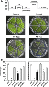
Phenotype characterization of PME T-DNA insertion lines under acquired thermotolerance (AT). Six-day-old seedlings of Arabidopsis wild type (Col), the HSP101 mutant (hsp101), and PME-defective (pme) lines were treated with 22°C for 2 h as a control and treated with 37°C, 1 h (sublethal HS [HS]) → 22°C, 2 h (recovery from HS; recovery) → 44°C, and 160 min (LHS) for the AT test. The pictogram shows the HS regime. A, Photographs were taken after 7 to 10 d from the end of treatment. B, Seedlings that survived were counted and survival rate was calculated (%). Two alleles of PME34-null mutants, pme34-1 and pme34-2, were hypersensitive toward the AT test. Data are means ± sd from three independent replicates (n = 150 seedlings). *, Significant at P < 0.05 compared with Col.
Figure 2.
Figure 2.
PME gene response to abiotic stresses and hormone treatments. A, The expression levels of PME34, PME28, and PME7 in response to abiotic stresses were analyzed by RT-PCR. Six-day-old seedlings were treated with 37°C HS for 3 h (HS) and then with HS with recovery at 22°C for 3 h (HS recovery), 150 mm NaCl for 8 h, as well as 6-h treatments of 300 mm d-mannitol, 0.5 mm H2O2, 250 μm NaAsO2 (As), and 500 μm CdCl2 (Cd). Treatment at 22°C with water was used as a control. The transcript level of PME34 was highly restored at the recovery phase after 37°C HS. B, The expression of PME34 was analyzed with the application of different concentrations of ABA, gibberellic acid (GA), and tert-butyl hydroperoxide (t-BH) as indicated. 18S rRNA was used as a loading control. C to E, Six-day-old Col seedlings were analyzed by basal (C) and acquired (D) thermotolerance tests as well as by different concentrations of ABA for 3 h (E). Gene expression was analyzed by qRT-PCR, and pictograms show the HS regime. The heat-responsive gene HSP18.1 (At5g59720) and the ABA-responsive gene RD29B (At5g52300) were used as references. The fold expression was normalized relative to 22°C control (CK) or water treatment. Data are means ± sd of three biological replicates. PP2AA3 (PP2A) was used as an internal control.
Figure 3.
Figure 3.
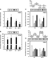
Transcriptional profiles of PME34 in ABA-deficient (aba2-1) and ABA-insensitive (abi1-1) plants in response to ABA and heat. Six-day-old seedlings of aba2-1 and abi1-1 were incubated in water containing 30 μm ABA for 3 h (A and B) and then analyzed by basal and acquired thermotolerance tests (C and D) as indicated in Figure 2. The expression levels of PME34 and RD29B were analyzed by qRT-PCR. The fold expression was normalized relative to their corresponding wild-type plants (Col or Ler) in water or 22°C control (CK) treatment. Data are means ± sd of three independent replicates. a, b, c, and * represent significantly different values compared with wild-type plants given water or 22°C control treatment (P < 0.05). PP2A was used as an internal control.
Figure 4.
Figure 4.
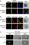
Subcellular localization analyses of PME34 in Arabidopsis mesophyll protoplasts and onion epidermal cells. A, PME34 was fused to N- or C-terminal GFP and coexpressed with the mCherry red fluorescent protein-labeled plasma membrane marker (RFP-PM) in protoplasts, as indicated. Blue shows chlorophyll using autofluorescent light. B, The protoplast fluorescence protease protection assay was performed with trypsin treatment. T-PMT, Transmitted light channel. C, Onion epidermal cells were treated with mannitol for the plasmolysis assay. GFP was used as a control. Bars = 20 μm.
Figure 5.
Figure 5.
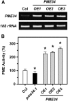
PME activity in PME34 overexpression lines. Six-day-old seedlings of Col, pme34-1, and three independent CaMV 35S promoter::GFP-PME34 lines (PME34-OE1, PME34-OE2, and PME34-OE3) were analyzed. A, PME34 expression levels were analyzed by RT-PCR. 18S rRNA was used as a loading control. B, PME activity (%) was normalized to that of Col. Data are means ± sd of three independent replicates. *, Significant at P < 0.05 compared with Col.
Figure 6.
Figure 6.
Thermotolerance test of pme34-1, PME34 overexpression (PME34-OE1, PME34-OE2, and PME34-OE3), PME3 complementation in a pme34-1 background (PME34-C3), and pme34-1 abi1-1 mutants. Acquired thermotolerance analysis was performed in 7-d-old seedlings, and pictograms show the HS regime. Plates were incubated without (A, B, and D) or with (C and E) 2.5 mL of 10 μm ABA for 12 h before heat at 37°C for 1 h. hsp101 was used as a reference. Survival was measured after 10 or 14 d from the end of treatment. Data are means ± sd of three biological replicates (n = 150 seedlings). *, Significant at P < 0.05 compared with wild-type plants, Col or Ler.
Figure 7.
Figure 7.
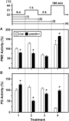
PME and PG activity in Col and pme34 plants in response to HS. Six-day-old seedlings of Col and pme34-1 were analyzed. The pictogram shows the HS regime. The activity (%) of PME (A) and PG (B) was normalized to the control (Col at 22°C treatment). Data are means ± sd of three independent replicates. *, Significant at P < 0.05 compared with Col.
Figure 8.
Figure 8.
Transcriptional levels and protein accumulations of major heat response genes in Col and pme34 plants in response to heat. Six-day-old seedlings of Col and pme34-1 were treated without (−) or with (+) heat at 37°C for 1 h (HS). A, The expression of the heat up-regulated marker genes HSP18.1, HSP70, HSP90, and HSP101 was analyzed by RT-PCR. B, The accumulation of class I small HSPs (sHSP-CI) and HSP70 was analyzed by western blotting. 18S rRNA and actin were used as loading controls.
Figure 9.
Figure 9.
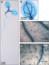
Histochemical analysis of PME34 promoter::GUS expression. Seven-day-old seedlings were analyzed for GUS activity. A and B, GUS expression of PME34 in a seedling (A) and cotyledon magnification of A (B). C and D, Cotyledons of the same seedling were treated without (C; −) or with (D; +) 30 μm ABA for 3 h, and then specimens were immersed together in staining solution.
Figure 10.
Figure 10.
Transpiration rate in Col and pme34 plants after mild heat treatment. A and B, Leaves of Col and pme34-1 plants were collected following treatment without (A; −) or with (B; +) heat at 37°C for 1 h. Transpiration rate was examined by measuring water loss (% of initial fresh weight) and recorded every 20 min for 120 min at room temperature (RT; 24°C–26°C and 55%–70% humidity), as indicated. The pictogram shows the HS regime. C, Merged data from A and B. Data are means ± sd of three independent replicates (n = 50 seedlings). *, Significant at P < 0.05 compared with Col.
Figure 11.
Figure 11.
Transpiration rate in Col and pme34 plants after lethal heat treatment. A to C, Leaves of Col and pme34-1 plants were collected following 30 (A), 80 (B), or 160 (C) min of 44°C lethal heat treatment, and then the transpiration rates were measured at room temperature (RT), as indicated in Figure 10. The pictogram shows the HS regime. The graphs at right depict the merged transpiration rates of Col and pme34-1 from A to C. Data are means ± sd of three independent replicates (n = 50 seedlings). *, Significant at P < 0.05 compared with Col.
Figure 12.
Figure 12.
Stomatal aperture in Col and pme34 plants after lethal heat treatment. A and B, Leaves of Col and pme34-1 plants were collected following 80 (A) or 160 (B) min of 44°C lethal heat treatment, and then stomatal apertures were recorded every 20 min at room temperature (RT), as indicated Figure 10. The pictogram shows the HS regime. Stomatal apertures were measured in terms of width-length ratio. Data are means ± sd of three independent replicates (n = 150 stomata). *, Significant at P < 0.05 compared with Col.

Similar articles

Cited by

References

    1. Alexandrov VY (1994) Functional aspects of cell response to heat shock. Int Rev Cytol 148: 171–227 - PubMed
    1. Atkinson RG, Schröder R, Hallett IC, Cohen D, MacRae EA (2002) Overexpression of polygalacturonase in transgenic apple trees leads to a range of novel phenotypes involving changes in cell adhesion. Plant Physiol 129: 122–133 - PMC - PubMed
    1. Baron KN, Schroeder DF, Stasolla C (2012) Transcriptional response of abscisic acid (ABA) metabolism and transport to cold and heat stress applied at the reproductive stage of development in Arabidopsis thaliana. Plant Sci 188-189: 48–59 - PubMed
    1. Bellincampi D, Camardella L, Delcour JA, Desseaux V, D’Ovidio R, Durand A, Elliot G, Gebruers K, Giovane A, Juge N, et al. (2004) Potential physiological role of plant glycosidase inhibitors. Biochim Biophys Acta 1696: 265–274 - PubMed
    1. Bethke G, Grundman RE, Sreekanta S, Truman W, Katagiri F, Glazebrook J (2014) Arabidopsis PECTIN METHYLESTERASEs contribute to immunity against Pseudomonas syringae. Plant Physiol 164: 1093–1107 - PMC - PubMed

MeSH terms