Review
. 2017 Jun;233(3):R131-R143.
doi: 10.1530/JOE-16-0679. Epub 2017 Apr 5.

Insulin receptor substrate signaling controls cardiac energy metabolism and heart failure

Affiliations
Review

Insulin receptor substrate signaling controls cardiac energy metabolism and heart failure

Cathy A Guo et al. J Endocrinol. 2017 Jun.

Abstract

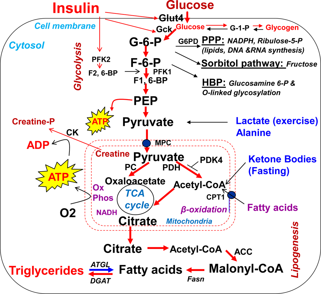
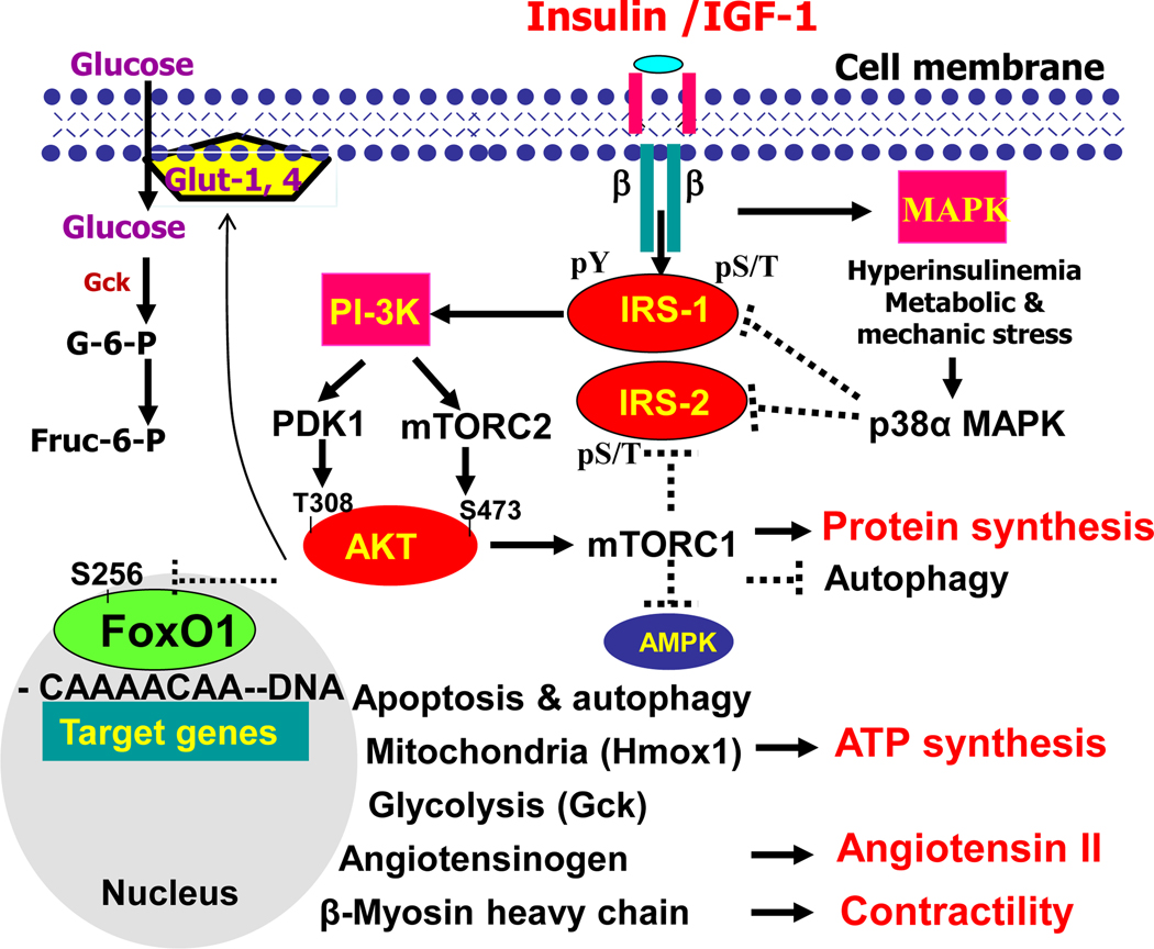
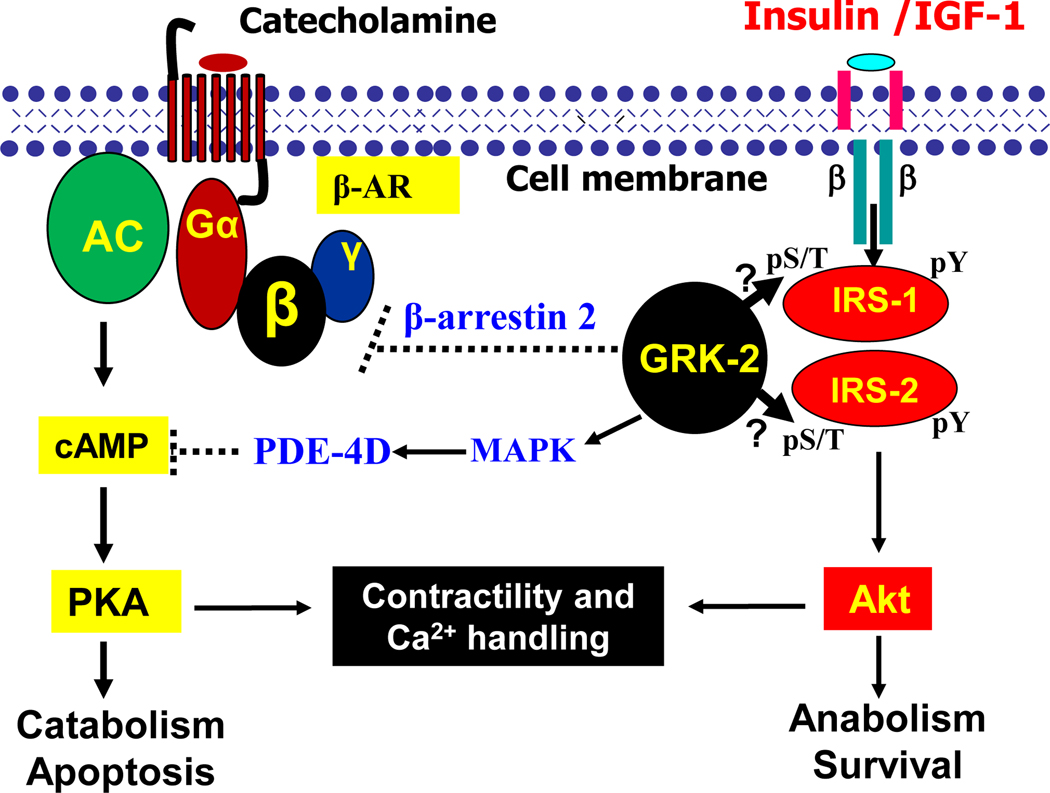
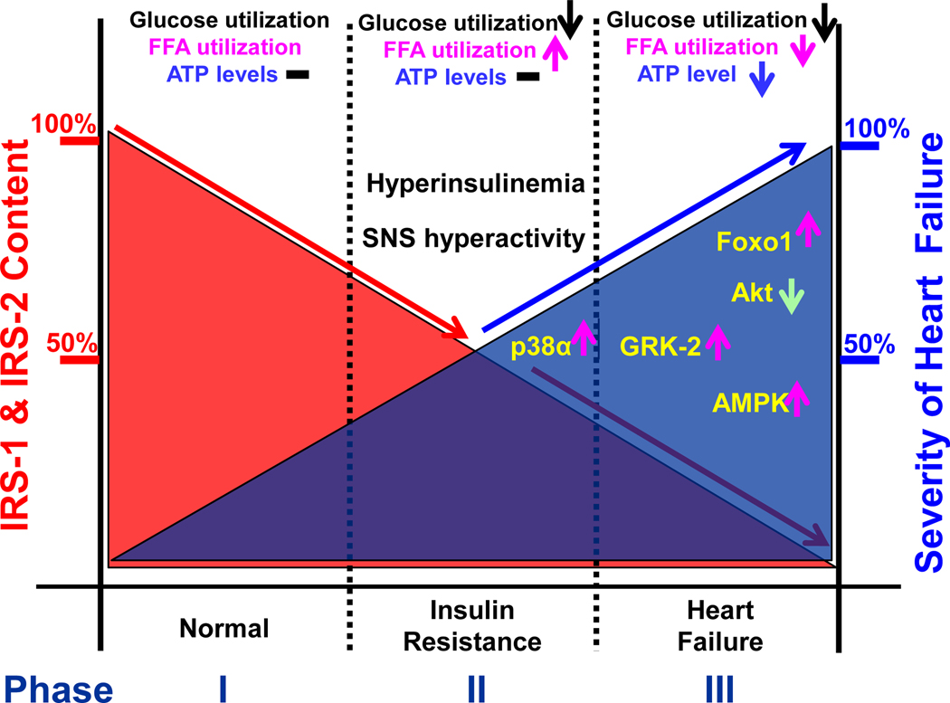
The heart is an insulin-dependent and energy-consuming organ in which insulin and nutritional signaling integrates to the regulation of cardiac metabolism, growth and survival. Heart failure is highly associated with insulin resistance, and heart failure patients suffer from the cardiac energy deficiency and structural and functional dysfunction. Chronic pathological conditions, such as obesity and type 2 diabetes mellitus, involve various mechanisms in promoting heart failure by remodeling metabolic pathways, modulating cardiac energetics and impairing cardiac contractility. Recent studies demonstrated that insulin receptor substrates 1 and 2 (IRS-1,-2) are major mediators of both insulin and insulin-like growth factor-1 (IGF-1) signaling responsible for myocardial energetics, structure, function and organismal survival. Importantly, the insulin receptor substrates (IRS) play an important role in the activation of the phosphatidylinositide-3-dependent kinase (PI-3K) that controls Akt and Foxo1 signaling cascade, regulating the mitochondrial function, cardiac energy metabolism and the renin-angiotensin system. Dysregulation of this branch in signaling cascades by insulin resistance in the heart through the endocrine system promotes heart failure, providing a novel mechanism for diabetic cardiomyopathy. Therefore, targeting this branch of IRS→PI-3K→Foxo1 signaling cascade and associated pathways may provide a fundamental strategy for the therapeutic and nutritional development in control of metabolic and cardiovascular diseases. In this review, we focus on insulin signaling and resistance in the heart and the role energetics play in cardiac metabolism, structure and function.

Keywords: forkhead transcription factor Foxo1; heart failure; insulin receptor substrates; insulin resistance.


Conflict of interest statement

Declaration of Interest

The author declares that there is no conflict of interest that could be perceived as prejudicing the impartiality of the review reported.

Figures

Figure 1.
Figure 1.
Metabolic flexibility of cardiomyocyte in use of glucose, fatty acids, lactate, amino acids, ketone bodies for generation of ATP to support cardiac contractile function. Insulin stimulates anabolic metabolism, including glucose uptake, glycolysis, and synthesis of glycogen, ribonucleotide, and lipid synthesis, while insulin inhibits the β-oxidation of fatty acids. An excess amount of ATP can be stored in creatine phosphate, and activated pentose phosphate pathway promotes synthesis of macromolecules and cardiac hypertrophy. Hexosamine biosynthetic pathway promotes glycosylation of many cellular proteins and bioactivity of target proteins and biological responses, particularly under hyperglycemia or insulin resistance. G-6-P-glucose-6-phosphate, G-1-P-gluocse-1-phosphate, F-6-P-fructose-6-phosphate, F-1,6-BP-fructose-1.2,-biphosphate, F2,6-BP-fructoase-2,6-biphosphate, PFK1-phosphofructokinase-1, PFK2-phosphofructokinase-2, NADPH-nicotinamide adenine dinucleotide phosphate, AR-aldose reductase, GFA-glutamine fructose-6-phosphate aminotransferase, PEP-phosphoenolpyruvate, MPC-mitochondrial pyruvate carrier, TCA-tricarboxylic acid, PPP-pentose phosphate pathway, G6PD-glucose-6-phosphate dehydrogenase, Gck-glucokinase, Glut- glucose transporter, HBP-hexosamine biosynthetic pathway, CTP1-carnitine-palmitoyltransferase-1, PDK4-pyruvate dehydrogenase kinase-4, PDH-pyruvate dehydrogenase, Ox phos-oxidative phosphorylation, β-oxidation-fatty acid beta-oxidation, NADH-nicotinamide adenine dinucleotide, DGAT-diacylglycerol O-acyltransferase, ATGL-adipose triglyceride lipase, Fasn-fatty acid synthase, TAG-triglycerides. ACC- Acetyl-CoA carboxylase, PC-pyruvate carboxylase, CK-Creatine kinase, Creatine-P- Creatine phosphate.
Figure 2.
Figure 2.
Insulin/IGF-1 signaling via IRS1,2-coupled PI-3K→AKT→Foxo1 and mTOR activation in control of energy metabolism, protein synthesis, mitochondrial function, autophagy, apoptosis, and gene expression of angiotensinogen and β-myosin heavy chain in cardiomyocytes. Insulin or IGF-1 binding to the insulin receptor or IGF-1 receptor, stimulates the receptor tyrosine kinases, recruiting IRS-1,−2 and activating PI-3K to generate second messenger PIP3, which then activates PDK1 and mTORC2 for the activation of downstream effectors, including Akt which promotes mTORC1 activity and protein synthesis and inactivation of AMPK and autophagy. In addition to promoting glut-1, −4 gene expression and their cellular membrane association, PI-3K and Akt also suppress transcription factor Foxo1, which promotes apoptosis and autophagy, gene expression of angiotensinogen and β-myosin heavy chain, heme oxygenase −1, while suppressing glucokinase gene expression for glucose oxidation and unitization. Hyperinsulinemia or other metabolic and mechanic stress activates intracellular protein kinases, such as p38α, promotes serine and threonine phosphorylation of IRS, inhibits tyrosine phosphorylation of IRS, and promotes IRS ubiquitination or degradation, and desensitizes insulin/IGF-1 signaling propagation for Akt activation. Abbreviation: IR- insulin receptor; IRS- insulin receptor substrate; pY-phosphorylated tyrosine; pS/T- phosphorylated serine or threonine. PI-3K-phosphatidylinositol (PI)-3-kinase; PDK1- phosphoinositide-dependent protein kinase-1; Foxo1-Forkhead/winged helix transcription factor O-class member 1; mTORC1-mammalian target of rapamycin complex-1; mTORC2-mammalian target of rapamycin complex-2; Hmoxo-1-heme oxygenase-1; MAPK-mitogen-activated protein kinase.
Figure 3.
Figure 3.
Crosstalk between insulin/IGF-1 signaling and β-adrenergic receptor signaling in control of cardiac catabolism and anabolism via the GRK2. Binding of catecholamine agonists to the G-protein coupled receptor (GCPR) that has seven transmembrane domains produces a conformational change in the GPCR, which promotes the binding of G-protein to the intracellular binding site on the receptor. The G-protein is heterotrimeric and activated Gα and Gβγ subunits are responsible for the activation of specific effectors, which produce different second messengers that generate a wide range of cellular responses, particularly for Ca2+ handling, cardiac contractility, catabolism, and apoptosis. The desensitization of GPCR is triggered by interaction with GRK-2 which phosphorylates the β-AR and enhances its interaction with β-arrestin. GRK-2 also activates PDE-4D gene expression by activating MAPK, thus suppressing cAMP production and PKA activity. An increase in receptor affinity toward β-arrestin for Gα protein uncoupling to Gβγ subunit which arrests the signal propagation. It is expected that GRK-2 may interact and phosphorylate IRS-1 and IRS-2, desensitizing insulin/IGF-1 signaling in the activation of PI-3K and Akt for control of anabolism and survival. Abbreviation: AC- adenylate cyclase; β-AR- beta-adrenergic receptors; Gα- G-protein α subunit; β- G-protein β subunit; γ- G-protein γ subunit; GRK-2- G-protein coupled receptor kinase-2; cAMP- cyclic 3’,5’-adenosine monophosphate; PDE-4D- phosphodiesterase 4D.
Figure 4.
Figure 4.
A proposed model for the dynamic changes of cardiac IRS-1 and IRS-2 synthesis in control of metabolic adaptation, cardiac insulin resistance, remodeling, and heart failure. The cardiac IRS-1 and IRS-2 protein levels are tightly correlated to the severity of heart failure and associated with myocardial intracellular protein kinases and Foxo1 activation. Under feeding conditions, IRS proteins slightly decreased to less than 25% and ATP homeostasis is maintained by glucose and free fatty acid (FFA) oxidation. Under insulin resistant state, further down regulation of IRS protein by less than 50% desensitizes glucose uptake and utilization with enhanced FFA oxidation, thus ATP homeostasis can be well maintained even when obesity occurs. However, down regulation of IRS proteins by more than 50% will result in cardiac dysfunction and 100% loss of IRS proteins will cause severe heart failure and death, in which a number of factors including activation of p38α, GRK-2, AMPK, and Foxo1, as well as inactivation of Akt are shown.

References

    1. Abel ED, Kaulbach HC, Tian R, Hopkins JC, Duffy JT, Minnemann T, Boers ME, Hadro E, Oberste-Berghaus C, et al. 1999. Cardiac hypertrophy with preserved contractile function after selective deletion of GLUT4 from the heart. J Clin Invest 104 1703–1714. - PMC - PubMed
    1. Anderson EA, Hoffman RP, Balon TW, Sinkey CA & Mark AL 1991. Hyperinsulinemia produces both sympathetic neural activation and vasodilation in normal humans. J Clin Invest 87 2246–2252. - PMC - PubMed
    1. Battiprolu PK, Gillette TG, Wang ZV, Lavandero S & Hill JA 2010. Diabetic Cardiomyopathy: Mechanisms and Therapeutic Targets. Drug Discov Today Dis Mech 7 e135–e143. - PMC - PubMed
    1. Battiprolu PK, Hojayev B, Jiang N, Wang ZV, Luo X, Iglewski M, Shelton JM, Gerard RD, Rothermel BA, Gillette TG, et al. 2012. Metabolic stress-induced activation of FoxO1 triggers diabetic cardiomyopathy in mice. J Clin Invest 122 1109–1118. - PMC - PubMed
    1. Belke DD, Betuing S, Tuttle MJ, Graveleau C, Young ME, Pham M, Zhang D, Cooksey RC, McClain DA, Litwin SE, et al. 2002. Insulin signaling coordinately regulates cardiac size, metabolism, and contractile protein isoform expression. J Clin Invest 109 629–639. - PMC - PubMed

Publication types