

Vertex-Specific Proteins pUL17 and pUL25 Mechanically Reinforce Herpes Simplex Virus Capsids
- PMID: 28381566
- PMCID: PMC5446649
- DOI: 10.1128/JVI.00123-17
Vertex-Specific Proteins pUL17 and pUL25 Mechanically Reinforce Herpes Simplex Virus Capsids
Abstract
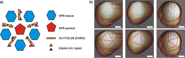
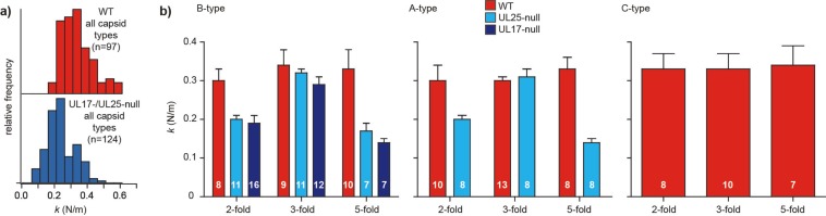
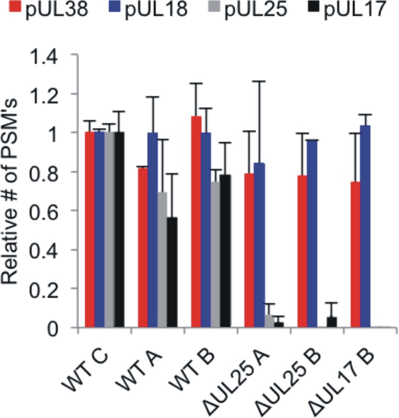
Using atomic force microscopy imaging and nanoindentation measurements, we investigated the effect of the minor capsid proteins pUL17 and pUL25 on the structural stability of icosahedral herpes simplex virus capsids. pUL17 and pUL25, which form the capsid vertex-specific component (CVSC), particularly contributed to capsid resilience along the 5-fold and 2-fold but not along the 3-fold icosahedral axes. Our detailed analyses, including quantitative mass spectrometry of the protein composition of the capsids, revealed that both pUL17 and pUL25 are required to stabilize the capsid shells at the vertices. This indicates that herpesviruses withstand the internal pressure that is generated during DNA genome packaging by locally reinforcing the mechanical sturdiness of the vertices, the most stressed part of the capsids.IMPORTANCE In this study, the structural, material properties of herpes simplex virus 1 were investigated. The capsid of herpes simplex virus is built up of a variety of proteins, and we scrutinized the influence of two of these proteins on the stability of the capsid. For this, we used a scanning force microscope that makes detailed, topographic images of the particles and that is able to perform mechanical deformation measurements. Using this approach, we revealed that both studied proteins play an essential role in viral stability. These new insights support us in forming a complete view on viral structure and furthermore could possibly help not only to develop specific antivirals but also to build protein shells with improved stability for drug delivery purposes.
Keywords: AFM; capsid; herpes simplex virus; stability.
Copyright © 2017 American Society for Microbiology.
Figures
Similar articles
-
Role of the Herpes Simplex Virus CVSC Proteins at the Capsid Portal Vertex.J Virol. 2020 Nov 23;94(24):e01534-20. doi: 10.1128/JVI.01534-20. Print 2020 Nov 23. J Virol. 2020. PMID: 32967953 Free PMC article.
-
Structure of the pseudorabies virus capsid: comparison with herpes simplex virus type 1 and differential binding of essential minor proteins.J Mol Biol. 2013 Sep 23;425(18):3415-28. doi: 10.1016/j.jmb.2013.06.034. Epub 2013 Jul 1. J Mol Biol. 2013. PMID: 23827137 Free PMC article.
-
The large tegument protein pUL36 is essential for formation of the capsid vertex-specific component at the capsid-tegument interface of herpes simplex virus 1.J Virol. 2015 Feb;89(3):1502-11. doi: 10.1128/JVI.02887-14. Epub 2014 Nov 19. J Virol. 2015. PMID: 25410861 Free PMC article.
-
Herpesvirus Capsid Assembly and DNA Packaging.Adv Anat Embryol Cell Biol. 2017;223:119-142. doi: 10.1007/978-3-319-53168-7_6. Adv Anat Embryol Cell Biol. 2017. PMID: 28528442 Free PMC article. Review.
-
Herpesvirus capsid assembly: insights from structural analysis.Curr Opin Virol. 2011 Aug;1(2):142-9. doi: 10.1016/j.coviro.2011.06.003. Curr Opin Virol. 2011. PMID: 21927635 Free PMC article. Review.
Cited by
-
Analytical Techniques to Characterize the Structure, Properties, and Assembly of Virus Capsids.Anal Chem. 2019 Jan 2;91(1):622-636. doi: 10.1021/acs.analchem.8b04824. Epub 2018 Dec 3. Anal Chem. 2019. PMID: 30383361 Free PMC article. Review.
-
Controlling the Revolving and Rotating Motion Direction of Asymmetric Hexameric Nanomotor by Arginine Finger and Channel Chirality.ACS Nano. 2019 Jun 25;13(6):6207-6223. doi: 10.1021/acsnano.8b08849. Epub 2019 May 28. ACS Nano. 2019. PMID: 31067030 Free PMC article. Review.
-
Structure of the herpes simplex virus 1 capsid with associated tegument protein complexes.Science. 2018 Apr 6;360(6384):eaao7298. doi: 10.1126/science.aao7298. Epub 2018 Apr 5. Science. 2018. PMID: 29622628 Free PMC article.
-
The interferon-inducible GTPase MxB promotes capsid disassembly and genome release of herpesviruses.Elife. 2022 Apr 27;11:e76804. doi: 10.7554/eLife.76804. Elife. 2022. PMID: 35475759 Free PMC article.
-
Universal Identification of Pathogenic Viruses by Liquid Chromatography Coupled with Tandem Mass Spectrometry Proteotyping.Mol Cell Proteomics. 2024 Oct;23(10):100822. doi: 10.1016/j.mcpro.2024.100822. Epub 2024 Jul 30. Mol Cell Proteomics. 2024. PMID: 39084562 Free PMC article.
References
Publication types
MeSH terms
Substances
Grants and funding
LinkOut - more resources
Full Text Sources
Other Literature Sources
Miscellaneous
