. 2017 Jun 9;91(13):e02418-16.
doi: 10.1128/JVI.02418-16. Print 2017 Jul 1.

Discovery of Novel Small-Molecule Inhibitors of LIM Domain Kinase for Inhibiting HIV-1

Affiliations

Discovery of Novel Small-Molecule Inhibitors of LIM Domain Kinase for Inhibiting HIV-1

Fei Yi et al. J Virol. .

Abstract

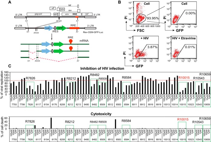
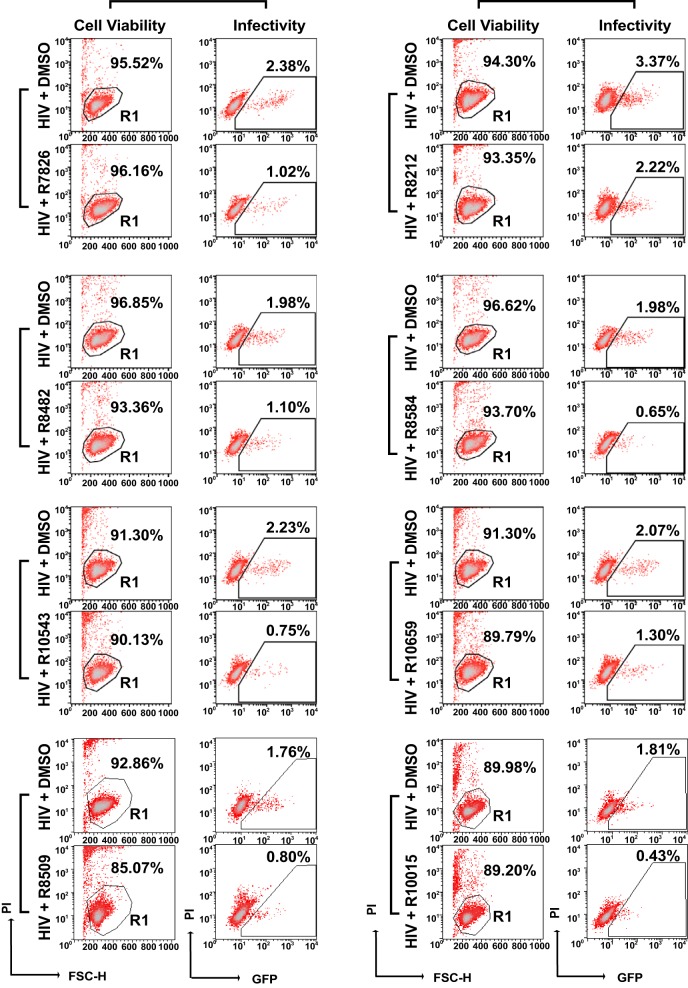
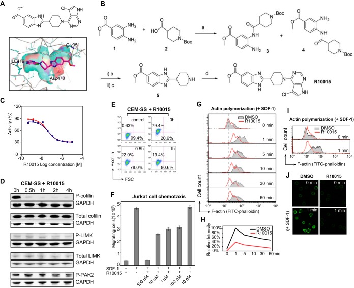
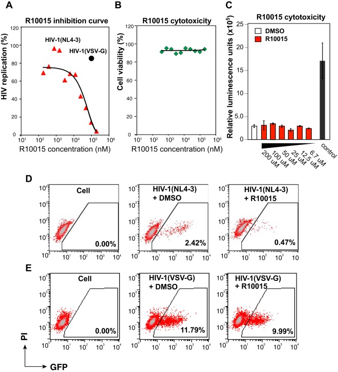
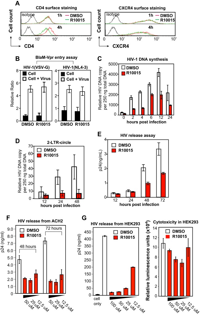
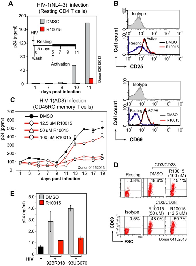
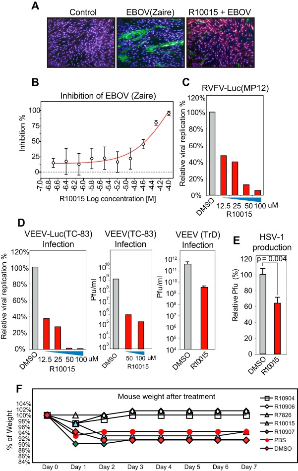
A dynamic actin cytoskeleton is necessary for viral entry, intracellular migration, and virion release. For HIV-1 infection, during entry, the virus triggers early actin activity by hijacking chemokine coreceptor signaling, which activates a host dependency factor, cofilin, and its kinase, the LIM domain kinase (LIMK). Although knockdown of human LIM domain kinase 1 (LIMK1) with short hairpin RNA (shRNA) inhibits HIV infection, no specific small-molecule inhibitor of LIMK has been available. Here, we describe the design and discovery of novel classes of small-molecule inhibitors of LIMK for inhibiting HIV infection. We identified R10015 as a lead compound that blocks LIMK activity by binding to the ATP-binding pocket. R10015 specifically blocks viral DNA synthesis, nuclear migration, and virion release. In addition, R10015 inhibits multiple viruses, including Zaire ebolavirus (EBOV), Rift Valley fever virus (RVFV), Venezuelan equine encephalitis virus (VEEV), and herpes simplex virus 1 (HSV-1), suggesting that LIMK inhibitors could be developed as a new class of broad-spectrum antiviral drugs.IMPORTANCE The actin cytoskeleton is a structure that gives the cell shape and the ability to migrate. Viruses frequently rely on actin dynamics for entry and intracellular migration. In cells, actin dynamics are regulated by kinases, such as the LIM domain kinase (LIMK), which regulates actin activity through phosphorylation of cofilin, an actin-depolymerizing factor. Recent studies have found that LIMK/cofilin are targeted by viruses such as HIV-1 for propelling viral intracellular migration. Although inhibiting LIMK1 expression blocks HIV-1 infection, no highly specific LIMK inhibitor is available. This study describes the design, medicinal synthesis, and discovery of small-molecule LIMK inhibitors for blocking HIV-1 and several other viruses and emphasizes the feasibility of developing LIMK inhibitors as broad-spectrum antiviral drugs.

Keywords: CXCR4; Ebola virus; LIM domain kinase; Rift Valley fever virus; Venezuelan equine encephalitis virus; actin; cofilin; cytoskeleton; herpes simplex virus; human immunodeficiency virus.


Figures

FIG 1
FIG 1
Screening LIMK inhibitors for anti-HIV activity. (A) Schematics of the Rev-dependent reporter construct and its transcripts in the HIV Rev-dependent indicator cell line Rev-CEM-GFP-Luc. The presence of Rev response element (RRE) in unspliced and singly spliced transcripts renders GFP/Luc expression Rev dependent. (B) Examples of HIV-dependent expression of GFP in Rev-CEM-GFP-Luc cells. Cells were not infected or infected with HIV(NL4-3) and treated with the reverse transcriptase inhibitor etravirine (100 nM). The cells were washed to remove the virus and the inhibitors and incubated for 48 h. HIV-dependent GFP expression was measured by flow cytometry. PI was added during flow cytometry to simultaneously measure cell viability. To exclude nonspecific cytotoxicity, only viable cells were used for GFP quantification. (C) Screening LIMK inhibitors with Rev-CEM-GFP-Luc cells. Cells were pretreated with LIMK inhibitors or DMSO for 1 h and then infected with HIV-1(NL4-3) for 3 h. The cells were washed to remove the virus and the inhibitors and incubated for 48 h. HIV-dependent GFP expression was measured by flow cytometry as described for panel B. PI was added during flow cytometry. The relative infection rates in drug-treated versus DMSO-treated cells (100%) were plotted using the relative percentage of GFP+ cells.
FIG 2
FIG 2
LIMK inhibitors discovered from anti-HIV screening. Rev-CEM-GFP-Luc HIV Rev-dependent indicator T cells were pretreated with LIMK inhibitors (100 μM) or DMSO for 1 h and then infected with HIV-1(NL4-3) for 3 h. The cells were washed to remove the virus and the inhibitors and incubated for 48 h. HIV-dependent GFP expression was measured by flow cytometry. PI was added during flow cytometry to simultaneously measure cell viability. Only viable cells (gated R1) were used for GFP quantification. The percentages of GFP+ cells are shown.
FIG 3
FIG 3
Chemical and biochemical characterization of R10015. (A) Chemical structure of R10015 and its docking into the crystal structure of LIMK1 (PDB accession no. 3S95, chain A). The binding motif of R10015 shows that it is a typical type I ATP-competitive kinase inhibitor. (B) R10015 synthesis. (a) EDC/HOBt/DIEA in DMF at room temperature for 16 h. (b) Acetic acid at 70°C for 4 h. (c) TFA (30%) in DCM at room temperature for 1 h. (d) 4,5-Dichloro-7H-pyrrolo[2,3-d]pyrimidine, DIEA, and isopropanol; 130°C; microwave for 3 h. (C) Ten-dose inhibition curve of R10015 against purified recombinant LIMK1 enzyme (IC50 = 38 ± 5 nM). (D) R10015 blocks cofilin serine 3 phosphorylation in human T cells. CEM-SS T cells were treated with R10015 (100 μM) for a time course, and the phosphorylation of cofilin, LIMK, and PAK2 was measured by Western blotting. GAPDH was used as a loading control. (E) Cofilin phosphorylation in R10015-treated (100 μM) CEM-SS T cells was also quantified by intracellular staining with an anti-p-cofilin antibody and analyzed by flow cytometry. The control was the background staining of cells in the absence of the anti-p-cofilin antibody. (F) R10015 inhibits Jurkat T cell chemotaxis in responding to SDF-1. Cells were treated with R10015 (100 μM) and added to the upper chamber of a 24-well transwell plate. The lower chamber was filled with SDF-1 (40 ng/ml). The plate was incubated at 37°C for 2 h, and the cells that migrated to the lower chamber were counted. FSC, forward scatter. The error bars indicate standard deviation. (G) R10015 inhibits chemotactic actin activity. R10015-treated (100 μM) resting CD4 T cells were exposed to SDF-1. Actin polymerization was measured by FITC-phalloidin staining. (H) The relative intensity of F-actin staining was also plotted. (I and J) Repeat of the experiment in panel H in another donor. Actin polymerization was quantified by flow cytometry (I) and confocal fluorescence microscopy (J).
FIG 4
FIG 4
R10015 inhibition of HIV infection of human T cells. (A) Rev-CEM-GFP-Luc cells were pretreated with different doses of R10015 for 1 h and then infected with HIV-1(NL4-3) for 3 h. The cells were washed to remove the virus and the inhibitor and incubated for 48 h. HIV-dependent luciferase expression was quantified, and the IC50 of R10015 was calculated (red triangles). For comparison, cells were also treated with 100 μM R10015 and infected with a VSV G-pseudotyped HIV for 3 h (black circle). Following infection, the cells were washed to remove the virus and R10015 and incubated for 48 h. (B) The cytotoxicity of R10015 was simultaneously measured by PI staining and flow cytometry. (C) The cytotoxicity of R10015 was also measured using a luciferase-based multiplex cytotoxicity assay, as described in Materials and Methods. For cytotoxicity control, 1% saponin was added to the cells to induce cytotoxicity (control). The error bars indicate standard deviations. (D and E) Flow cytometry results demonstrating that R10015 inhibited HIV-1(NL4-3) but minimally inhibited HIV-1(VSV G) when the cells were briefly treated with R10015 (100 μM) early during viral infection.
FIG 5
FIG 5
R10015 inhibits HIV-1 DNA synthesis, nuclear migration, and virion release. (A) Effects of R10015 on surface CD4 and CXCR4 expression. CEM-SS T cells were treated with R10015 (100 μM) and then stained for surface CD4 or CXCR4. (B) R10015 did not inhibit viral entry. CEM-SS T cells were treated with R10015 (100 μM) and then infected with BlaM-Vpr-tagged HIV-1(NL4-3) or HIV-1(VSV G) to measure viral entry. (C) R10015 inhibits viral DNA synthesis. CEM-SS T cells were treated with R10015 (100 μM) for 1 h and then infected with a single-cycle HIV-1(Env) for 2 h in the presence of R10015. Following infection, the cells were washed to remove HIV-1 and R10015. Viral DNA synthesis was measured by real-time PCR. (D) R10015 inhibits 2-LTR circle DNA formation. Cells were similarly treated with R10015 and infected. 2-LTR circles were quantified by real-time PCR. (E) R10015 inhibits virion release. Cells were infected with HIV-1(Env) for 2 h, washed, incubated for 12 h, and then treated with R10015. Virion release was quantified by measuring p24 in the supernatant. (F) R10015 inhibits virion release from chronically infected ACH2 cells. ACH2 cells were washed 3 times and then treated with R10015 at various dosages. The cells were cultured for 2 to 3 days. Culture supernatants were harvested and analyzed for HIV-1 p24 by ELISA. DMSO was used as a control. (G) R10015 inhibits virion release from DNA-transfected HEK293 cells. The cells were transfected with plasmid pHIV-1(NL4-3) and then treated with different concentrations of R10015 for 48 h. Culture supernatants were harvested and analyzed for HIV-1 p24 by ELISA. DMSO was used as a control. The cytotoxicity of R10015 in HEK293 cells was also measured using a luciferase-based multiplex cytotoxicity assay, as described in Materials and Methods. The error bars indicate standard deviations.
FIG 6
FIG 6
R10015 inhibits R5 and X4 HIV-1 latent infection of resting CD4 T cells and primary isolate infection of PBMC. (A) R10015 inhibits HIV latent infection of resting CD4+ T cells. Cells were treated with R10015 (100 μM) or DMSO for 1 h and infected with HIV-1(NL4-3) for 2 h. The virus and the drug were washed away, and the cells were cultured for 5 days in the absence of R10015 and then activated with CD3/CD28 beads. Viral p24 release was measured. (B) CD25 and CD69 surface staining demonstrates that R10015 did not inhibit T cell activation with this short period of drug treatment. (C) R10015 inhibits R5 HIV-1 latent infection of CD45RO+ memory CD4 T cells. Resting memory CD4 T cells were similarly treated with R10015, infected with HIV-1(AD8), washed, incubated for 5 days without stimulation, and then activated with CD3/CD28 beads. (D) CD69 surface staining was performed for control of R10015 effects on T cell activation. (E) R10015 inhibits HIV-1 primary isolate infection of PBMC. PBMC were cultured for 1 day and then treated with 100 μM R10015 for 1 h. The cells were infected with HIV 92/BR/018 (Brazil) or HIV 93UG070 (Uganda) for 3 h, washed to remove the viruses and R10015, and cultured for 3 days. The supernatant was analyzed for HIV-1 p24 by ELISA. DMSO was used as a control. The error bars indicate standard deviations.
FIG 7
FIG 7
R10015 inhibition of EBOV, VEEV, RVFV, and HSV-1. (A and B) Inhibition of EBOV by R10015. (A) HFF-1 cells were treated with R10015 (50 μM) for 2 h and infected with EBOV (Zaire) (MOI, 2.5) for 48 h in the presence of R10015. The cells were fixed and stained for the EBOV GP protein with an Alexa 488-labeled antibody (green) or with Hoechst (blue nuclei) for confocal imaging. (B) The relative GP protein staining was converted to percent inhibition using the infected, non-drug-treated cells as the control. (C) Inhibition of RVFV by R10015. Vero cells were similarly treated with R10015, infected with RVFV-Luc (MP12) (MOI, 0.1), and analyzed by luciferase assay. (D) Inhibition of VEEV by R10015. Vero cells were similarly treated with R10015 and infected with VEEV-Luc(TC-83), VEEV(TC-83), or VEEV(TrD) (MOI, 0.1). The viral supernatants were collected at 24 h and analyzed by luciferase or plaque assay. (E) Inhibition of HSV-1 by R10015. Vero cells were treated with R10015 (100 μM) for 2 h, infected with HSV-1, washed, and cultured in the absence of R10015. Viral plaques were stained and quantified. No drug toxicity was observed up to 100 μM R10015. (F) C3H/HeN mice were treated daily with LIMK inhibitors for 7 days. R10904, R10906, R10907, and R7826 were delivered via oral gavage at 20 mg/kg. R10015 was delivered by intraperitoneal injection at 10 mg/kg. DMSO-treated and PBS-treated mice were used as controls. The animals were weighed daily and observed for signs of stress. The error bars indicate standard deviations.

References

    1. Moore RD, Chaisson RE. 1999. Natural history of HIV infection in the era of combination antiretroviral therapy. AIDS 13:1933–1942. doi:10.1097/00002030-199910010-00017. - DOI - PubMed
    1. Nowak M. 1990. HIV mutation rate. Nature 347:522. - PubMed
    1. Arhel N, Kirchhoff F. 2010. Host proteins involved in HIV infection: new therapeutic targets. Biochim Biophys Acta 1802:313–321. doi:10.1016/j.bbadis.2009.12.003. - DOI - PubMed
    1. Spear M, Guo J, Wu Y. 2013. Novel anti-HIV therapeutics targeting chemokine receptors and actin regulatory pathways. Immunol Rev 256:300–312. doi:10.1111/imr.12106. - DOI - PMC - PubMed
    1. Jones KL, Smyth RP, Pereira CF, Cameron PU, Lewin SR, Jaworowski A, Mak J. 2011. Early events of HIV-1 infection: can signaling be the next therapeutic target? J Neuroimmune Pharmacol 6:269–283. doi:10.1007/s11481-011-9268-5. - DOI - PubMed

Publication types

MeSH terms