Review
. 2017 Jun 1;24(6):539-551.
doi: 10.5551/jat.40352. Epub 2017 Apr 4.

Recent Advances in Fluorescent Angioscopy for Molecular Imaging of Human Atherosclerotic Coronary Plaque

Affiliations
Review

Recent Advances in Fluorescent Angioscopy for Molecular Imaging of Human Atherosclerotic Coronary Plaque

Yasumi Uchida. J Atheroscler Thromb. .

Abstract

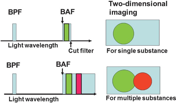
Purpose of review: In vivo imaging of the native substances, including lipoproteins, that comprise human atherosclerotic plaques is currently beyond the scope of any available imaging techniques. Color and near-infrared fluorescent angioscopy (CFA and NIRFA, respectively) systems have been recently developed for molecular imaging of lipoproteins within the human coronary arterial wall ex vivo and/or in vivo. The author reviews recent findings on lipoprotein deposition in human coronary plaques obtained by these imaging techniques.

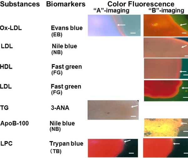
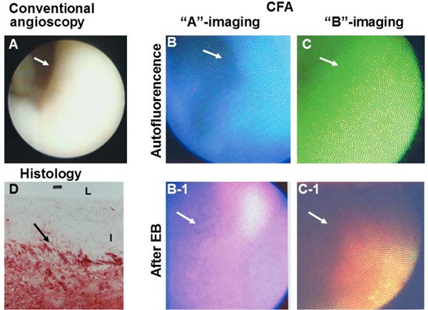
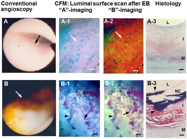
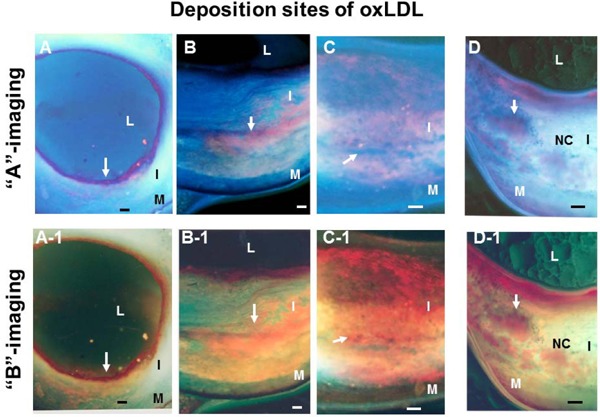
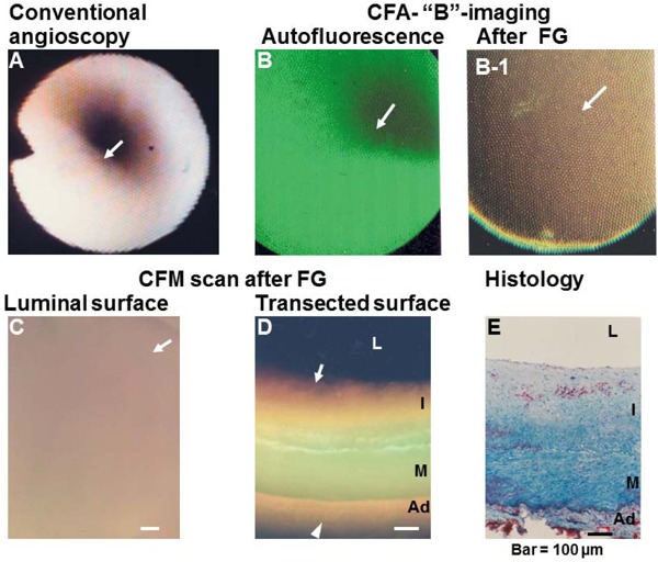
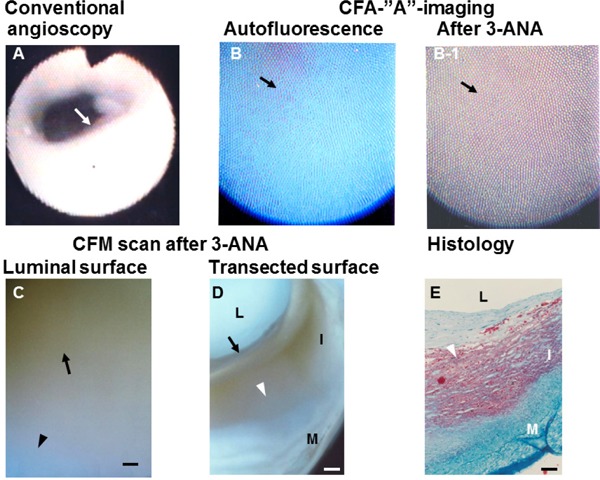
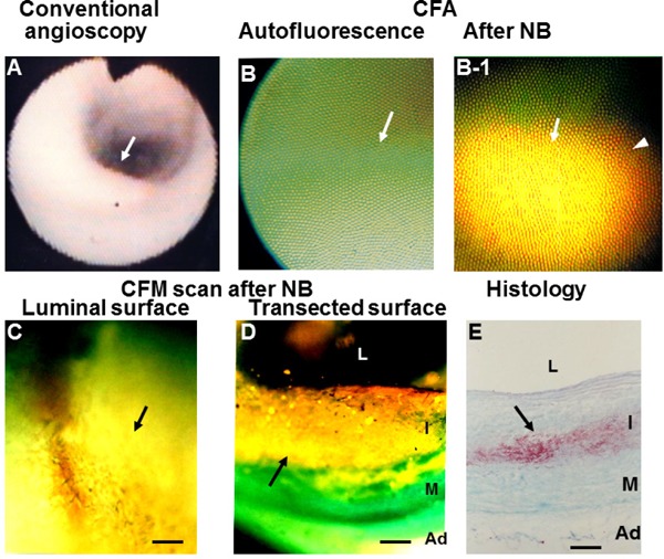
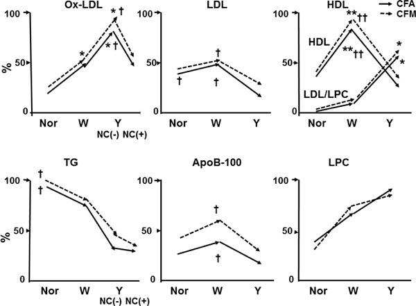
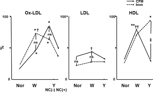
Recent findings: Using specific biomarkers, native pro-atherogenic substances such as oxidized low-density lipoprotein (ox-LDL), LDL, triglycerides (TG), apolipoprotein B-100 (ApoB-100), and lysophosphatidylcholine (LPC), and the anti-atherogenic substance such as high-density lipoprotein (HDL) were visualized by CFA, and LDL and cholesterol by NIRFA, in coronary plaques obtained from autopsy subjects. The relationship between incidence and plaque morphology differed for each substance. The incidence of ox-LDL and LDL on color fluorescence microscopy correlated well with that observed using immunohistochemical techniques. During coronary catheterization in patients, ox-LDL, LDL, and HDL in coronary plaques were visualized by CFA or NIRFA.

Conclusions: Using CFA or NIRFA, the distribution of the major native pro-atherogenic and anti-atherogenic lipoproteins and their components within human coronary plaques can be evaluated ex vivo and/or in vivo. Fluorescent angioscopy could help our understanding of the molecular mechanisms of coronary atherosclerosis and in the evaluation of the effects of therapy targeting the substances comprising atherosclerotic coronary plaques.

Keywords: Apolipoproteins; Biomarkers; Color fluorescent angioscopy; Immunohistochemistry; Lipoproteins; Near-infrared fluorescent angioscopy.


Figures

Fig. 1.
Fig. 1.
Schematic Representation of Fluorescent Imaging Techniques A light with a desired wave length is formed by passing white light through a band-pass filter (BPF) to radiate the target substance, and fluorescence that is exhibited by the target substance is selected through a band-absorption filter (BAF). A cut filter is set behind the BAF to image the fluorescence of the target substance alone (upper half), but the cut filter is not set to image fluorescence of both the target substance and other substances (lower half).
Fig. 2.
Fig. 2.
Fluorescent Colors of Major Substances Comprising Atherosclerotic Plaques Elicited by Biomarkers 3-ANA; 3-amino-4-hydroxy-5-nitrobenzenesulfonic acid monohydrate, ApoB-100; apolipoprotein B-100, HDL; high-density lipoprotein, LDL; low-density lipoprotein, LPC; lysophosphatidylcholine, Ox-LDL; oxidized low-density lipoprotein, TG; triglycerides. Bar = 100 µm.
Fig. 3.
Fig. 3.
Oxidized Low-density Lipoprotein (Ox-LDL) Imaged by Color Fluorescent Angioscopy (CFA) Using Evans Blue (EB) as a Biomarker White plaque imaged by conventional angioscopy. Arrow; portion observed by CFA. Before the administration of EB, the plaque shows blue auto-fluorescence with “A” imaging (B) and green auto-fluorescence with “B” imaging (C), indicating collagen I. After the intracoronary administration of EB, the plaque shows violet fluorescence with “A” imaging (B-1) and reddishbrown fluorescence (C-1), which indicates the deposition of ox-LDL. Lipids, which were stained with Oil Red-O and methylene blue, were deposited in deep layer but not in superficial layer of the plaque (D). L and I; lumen and intima, respectively. (cited with permission from ref. 25)
Fig. 4.
Fig. 4.
Oxidized Low-density Lipoprotein (Ox-LDL) Imaged by Luminal Surface Scan with Color Fluorescent Microscopy (CFM) A light-yellow plaque on conventional angioscopy (arrow in A) exhibited diffuse violet (arrow in A-1) and reddish-brown fluorescence (arrow in A-2) after the administration of EB, indicating the deposition of ox-LDL. Histology showed no obvious lipid deposition in the observed portion (A-3). A yellow plaque with a NC on conventional angioscopy and histology (arrows in B and B-3) exhibited web-like violet (arrow in B-1) or reddish-brown fluorescence (arrow in B-2) after the administration of EB, indicating deposition of ox-LDL. L, I, M, and NC; lumen, intima, media, and necrotic core, respectively. Bar: 100 µm. (Cited with permission from ref. 33)
Fig. 5.
Fig. 5.
Oxidized Low-density Lipoprotein (Ox-LDL) Imaged by Scanning the Transected Surface of Plaques by Color Fluorescent Microscopy (CFM) Ox-LDL deposited either in superficial layer (arrows in A, A-1), deep layer (arrows in B, B-1), or in both (arrows in C, C-1). Ox-LDL deposits were disseminated in the NC (arrows in D, D-1). L, I, M, and NC; lumen, intima, media, and necrotic core, respectively. Bar = 100 µm. (Cited with permission from ref. 33)
Fig. 6.
Fig. 6.
Deposition of High-density Lipoprotein (HDL) in a Coronary Plaque Visualized by “B” Imaging of Color Fluorescent Angioscopy (CFA) and Microscopy (CFM) (A) Normal coronary segment. Arrow; portion observed by CFA. (B) CFA image of the plaque in (A), showing green fluorescence that indicates collagen I (arrow). (B-1) CFA image after the administration of Fast green (FG), showing brown fluorescence that indicates the deposition of HDL (arrow). (C, D) CFM images of the luminal and transected surfaces of the same plaque, showing brown fluorescence that indicates HDL (arrow). (E) Histology of the same segment after Oil Red-O and methylene blue staining. Lipids (cholesterol and/or cholesteryl esters) show a spotty pattern of distribution in the deep layer of the intima (arrow). L, I, M, and Ad: lumen, intima, media, and adventitia, respectively. Bar = 100 µm.
Fig. 7.
Fig. 7.
Triglyceride (TG) Imaged by “A” Imaging of Color Fluorescent Angioscopy (CFA) Using 3-ANA as A Biomarker (A) White plaque (arrow). (B) Blue fluorescence of the same plaque imaged by CFA before the application of 3-ANA, indicating rich collagen I. (B-1) Dark-brown fluorescence elicited by the application of 3-ANA to the same plaque (arrow). (C). Color fluorescent microscopy (CFM) shows dark -brown luminal surface, indicating TG. The transected surface of the same plaque shows deposition of TG in the inner portions of plaque where lipid deposition was not observed (arrow in D). Arrowhead in (D) shows the lipid-laden portion in (E)(E) L, I, and M: lumen, intima, and media, respectively. Scale bars = 100 µm. (Cited with permission from ref. 39)
Fig. 8.
Fig. 8.
Apolipoprotein B-100 (ApoB-100) in a Coronary Plaque Imaged by Color Fluorescent Angioscopy (CFA) and Microscopy (CFM) White plaque. Arrow; section observed by CFA. (B) Same plaque before administration of Nile blue (NB) dye (arrow) shows green fluorescence, indicating collagen I. (B-1) Same plaque after administration of NB shows golden fluorescence, indicating diffuse deposition of ApoB-100 (arrow). (C) Luminal surface of the same plaque scanned by CFM shows diffuse and golden fluorescence (arrow). (D) Scan of the transected surface of the same plaque, showing diffuse deposition of ApoB-100 (arrow). (E) Histology of the same plaque after Oil Red-O and methylene blue dye staining shows deposition of lipids in deeper layer of the intima. L, I, M, and Ad: lumen, intima, media, and adventitia, respectively. Bar = 100 µm. (Cited with permission from ref. 36)
Fig. 9.
Fig. 9.
Relationship Between Plaque Morphology and the Percentage (%) Incidence of Lipoproteins and Their Components in Human Coronary Plaques Studied by Color Fluorescent Angioscopy (CFA) and Microscopy (CFM) Constructed using data from our studies [25, 27, 33, 35, 36, 37, 38, 39]. The autopsy subject group differed for substances examined. *; p < 0.05, **; p < 0.01 vs. normal segments (Nor). ; p < 0.05, ††; p < 0.01 vs. yellow plaques as a whole (Y) or Y with necrotic core [NC (+)]. No significant difference between CFA and CFM in any substances examined. Oxidized low-density lipoprotein (ox-LDL) increased in white plaques (W) and further in Y without [NC (−)] but decreased in Y NC (+); LDL decreased in Y (both NC (−) and NC (+); high-density lipoprotein (HDL) increased in W but decreased in Y, whereas LDL/LPC increased in Y; triglycerides (TG) decreased in W and further decreased in Y; ApoB-100 decreased in Y; and lysophosphatidylcholine (LPC) showed a tendency to increase in W and Y.
Fig. 10.
Fig. 10.
Comparison of the Percentage (%) Incidence of Lipoproteins Between Color Fluorescent Microscopy (CFM) and Immunohistochemical Staining (Imm) Constructed using data from our studies [25, 35, 37, 38, 43]. Autopsy subject groups differed between CFM and Imm. *; p < 0.05, **; p < 0.01 vs. normal segments. ; p < 0.05, p < 0.01. †† vs. yellow plaques as a whole (Y) or Y with necrotic core [NC (+)]. ; p < 0.05 between CFM and Imm. ns; not significant. The relationship between the incidence of oxidized low-density lipoprotein (ox-LDL) and plaque morphology did not differ between CFA and Imm. The incidence of low-density lipoprotein (LDL) was low for both CFM and Imm and showed no significant difference between them. High-density lipoprotein (HDL) increased in white plaques (W) and decreased in Y on CFM, whereas it increased further in Y on Imm. Because Fast green (FG) elicits a brown fluorescence of HDL while simultaneously eliciting red fluorescence of LDL and lysophosphatidylcholine (LPC), and because LDL/LPC increased in Y, it was considered that the brown fluorescence of HDL was masked by the red fluorescence of LDL/LPC, showing an apparent decrease in HDL in Y on CFM. ns; not significant.
Fig. 11.
Fig. 11.
Low-density Lipoprotein (LDL) Imaged by Near-infrared Fluorescent Angioscopy (NIRFA) Using Indocyanine Green (ICG) as a Biomarker in a Patient with Stable Angina (A) Angiogram of right coronary artery. (B) Conventional angioscopic (a-1–d-1) and NIRFA images (a-2–d-2) correspond to a–d in (A). Yellow plaque (a-1) and white plaque (c-1) exhibit NIRF. Arrows indicate the same portions observed by conventional angioscopy and NIRFA. (Cited with permission from ref. 42)

References

    1. Matsuura E, Hughes GR, Khamashta MA: Oxidation of LDL and its clinical implication. Autoimmun Rev, 2008; 7: 558-566 - PubMed
    1. Chen JH, Riazzy, Smith EM, Proud CG, Steinbrecher U: Oxidized LDL-mediated macrophage survival involves elongation of factor-2 kinase. Arterioscler Thromb Vasc Biol, 2009; 29: 92-98 - PubMed
    1. Tardif JC: Emerging high-density lipoprotein infusion therapies: fulfilling the promise of epidemiology? J Clin Lipidol, 2010; 4: 399-404 - PubMed
    1. Negi S, Ballantyne CM: Insights from recent meta-analysis: role of high-density lipoprotein cholesterol in reducing cardiovascular events and rates of atherosclerotic disease progression. J Clin Lipidol, 2010; 4: 365-370 - PubMed
    1. Vilani SS, Nambi V: The role of lipoprotein-associated phospholipse A2 as a marker of atherosclerosis. Curr Atheroscler Rep, 2007; 9: 97-103 - PubMed

MeSH terms