. 2017 Mar;20(1):20-26.
doi: 10.4048/jbc.2017.20.1.20. Epub 2017 Mar 24.

Inhibition of Nicotinamide Phosphoribosyltransferase Induces Apoptosis in Estrogen Receptor-Positive MCF-7 Breast Cancer Cells

Affiliations

Inhibition of Nicotinamide Phosphoribosyltransferase Induces Apoptosis in Estrogen Receptor-Positive MCF-7 Breast Cancer Cells

Mohammad Alaee et al. J Breast Cancer. 2017 Mar.

Abstract

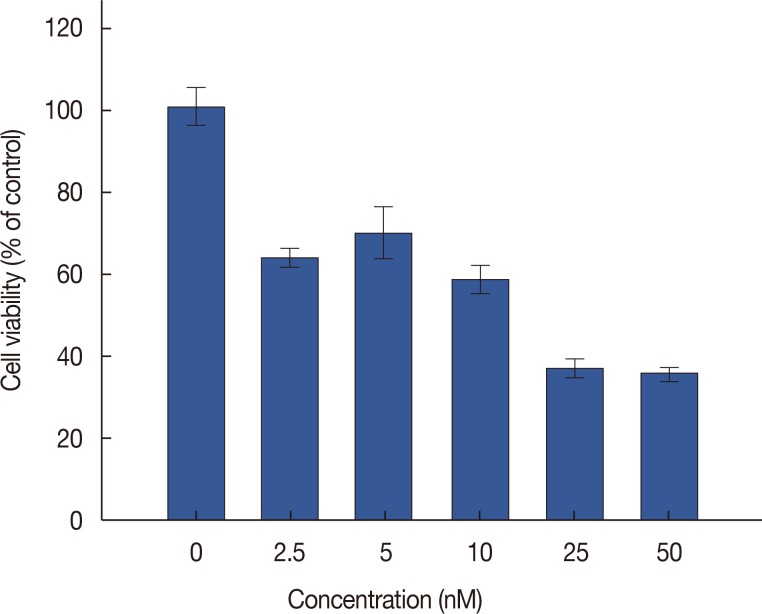
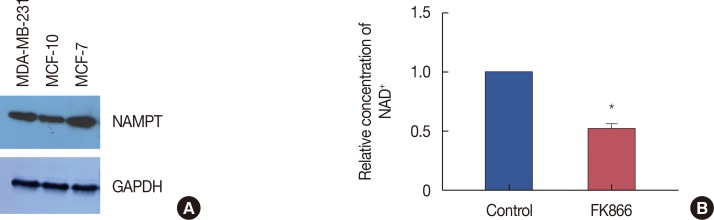
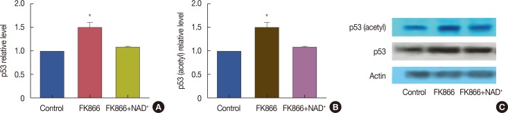
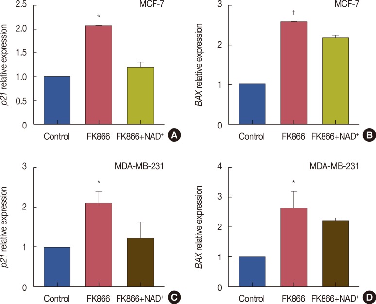
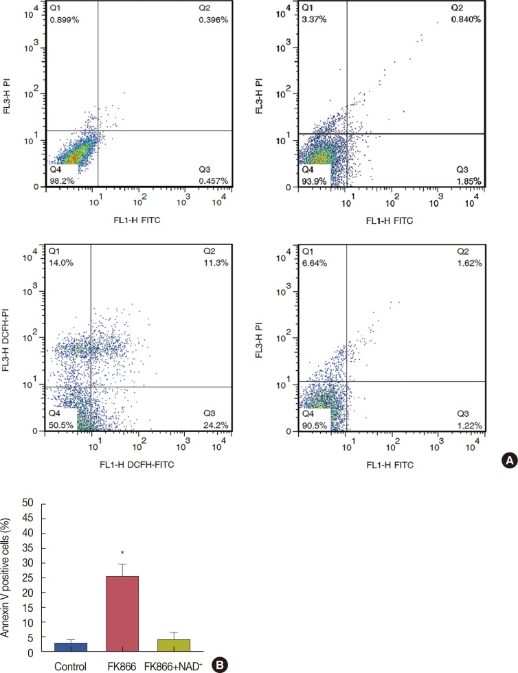
Purpose: Tumor cells have increased turnover of nicotinamide adenine dinucleotide (NAD+), the main coenzyme in processes including adenosine diphosphate-ribosylation, deacetylation, and calcium mobilization. NAD+ is predominantly synthesized in human cells via the salvage pathway, with the first component being nicotinamide. Nicotinamide phosphoribosyltransferase (NAMPT) is the key enzyme in this pathway, and its chemical inhibition by FK866 has elicited antitumor effects in several preclinical models of solid and hematologic cancers. However, its efficacy in estrogen receptor (ER)-positive and human epidermal growth factor receptor 2-positive breast cancer cells has not been previously investigated. In this study, we aimed to deplete the NAD+ content of MCF-7 cells, a model cell line for ER-positive breast cancer, by inhibiting NAMPT in order to evaluate downstream effects on p53 and its acetylation, p21 and Bcl-2-associated X protein (BAX) expression, and finally, apoptosis in MCF-7 breast cancer cells.

Methods: MCF-7 cells were cultured and treated with FK866. NAD+ levels in cells were determined colorimetrically. Levels of p53 and its acetylated form were determined by Western blotting. Expression of p21 and BAX was determined by real-time polymerase chain reaction. Finally, levels of apoptosis were assessed by flow cytometry using markers for annexin V and propidium iodide.

Results: FK866 treatment was able to increase p53 levels and acetylation, upregulate BAX and p21 expression, and induce apoptosis in MCF-7 cells. Addition of exogenous NAD+ to cells reversed these effects, suggesting that FK866 exerted its effects by depleting NAD+ levels.

Conclusion: Results showed that FK866 could effectively inhibit NAD+ biosynthesis and induce programmed cell death in MCF-7 cells, suggesting that NAMPT inhibitors may be useful for the treatment of ER-positive breast cancers.

Keywords: Apoptosis; Breast neoplasms; NAD; Nicotinamide phosphoribosyltransferase; Tumor suppressor protein p53.


Conflict of interest statement

CONFLICT OF INTEREST: The authors declare that they have no competing interests.

Figures

Figure 1
Figure 1. Cell viability assay to examine the cytotoxic effects of FK866 in MCF-7 cells. Cells were treated with various concentrations of FK866 (2.5–50 nM). The values of cell viability are represented as percent of control and each bar represents mean±standard error of the mean of six replicates in three separate experiments.
Figure 2
Figure 2. Nicotinamide phosphoribosyltransferase (NAMPT) expression and nicotinamide adenine dinucleotide (NAD) production. (A) NAMPT levels in MCF-7 cell compared to MCF-10A and MDA-MB-231 cells. (B) Relative concentration of NAD+ in treated cells compared to control. FK866 significantly decreased the NAD+ levels. MCF-7 cells were treated for 72 hours with 10 nM concentration of FK866 and NAD+ levels of treated and control cells were determined. Values are the mean of at least three independent experiments and are expressed as relative to control.
GAPDH=glyceraldehyde 3-phosphate dehydrogenase. *p<0.001.
Figure 3
Figure 3. Elevation of p53 and its acetylation after treatment with FK866 and nicotinamide adenine dinucleotide (NAD+). Cells were treated for 72 hours with FK866 (10 nM) or FK866+NAD+ (10 µM). (A) p53 protein levels in treated and control cells; (B) acetylated-p53 levels in treated and control cells; and (C) Western blotting evaluation of the effects of FK866 on p53 and its acetylated form. This experiment was repeated three times. Data from one representative experiment is shown. *p<0.05 compared to control.
Figure 4
Figure 4. FK866 induced p21 and BAX expression in MCF-7 and MDA-MB-231 cells. Cells were treated with FK866 alone or a combination of FK866 and nicotinamide adenine dinucleotide (NAD+) and gene expression of p21 and BAX was determined using real-time polymerase chain reaction. (A) Relative expression of p21 in treated MCF-7 cells compared to control; (B) relative expression of BAX in treated MCF-7 cells compared to control; (C) relative expression of p21 in treated MDA-MB-231 cells compared to control; (D) relative expression of BAX in treated MDA-MB-231 cells compared to control. Data are the mean of at least three independent experiments. *p<0.01; p<0.001.
Figure 5
Figure 5. FK866 caused apoptosis in MCF-7 cells while nicotinamide adenine dinucleotide (NAD+) reversed FK866-induced apoptosis. (A) Representative quadrant dot plot of annexin V-fluorescein isothiocyanate (FITC)/propidium iodide (PI) staining of MCF-7 cells measured by flow cytometry. 3×10⁵ Cells were seeded and treated with 10 nM FK866 for 36 hours. After incubation, cells were detached with accutase enzyme and after staining with annexin V and PI, were analyzed by flowcytometry. Separate regions of the plot are indicative of population of viable (annexin V−, PI−), early apoptotic (annexin V+, PI−), late apoptotic (annexin V+, PI+) and necrotic (annexin V−, PI+) cells. (B) Quantification of annexin V-positive cells indicating early apoptosis, as percentage of total cells. Data are representative of at least three independent experiments with similar results. *p<0.001.

References

    1. Chiarugi A, Dölle C, Felici R, Ziegler M. The NAD metabolome: a key determinant of cancer cell biology. Nat Rev Cancer. 2012;12:741–752. - PubMed
    1. Houtkooper RH, Auwerx J. Exploring the therapeutic space around NAD+ J Cell Biol. 2012;199:205–209. - PMC - PubMed
    1. Nikiforov A, Kulikova V, Ziegler M. The human NAD metabolome: functions, metabolism and compartmentalization. Crit Rev Biochem Mol Biol. 2015;50:284–297. - PMC - PubMed
    1. Garten A, Schuster S, Penke M, Gorski T, de Giorgis T, Kiess W. Physiological and pathophysiological roles of NAMPT and NAD metabolism. Nat Rev Endocrinol. 2015;11:535–546. - PubMed
    1. Hasmann M, Schemainda I. FK866, a highly specific noncompetitive inhibitor of nicotinamide phosphoribosyltransferase, represents a novel mechanism for induction of tumor cell apoptosis. Cancer Res. 2003;63:7436–7442. - PubMed