

Evaluation of the Prevalence of Maxillary Sinuses Abnormalities through Spiral Computed Tomography (CT)
- PMID: 28382118
- PMCID: PMC5375949
- DOI: 10.1055/s-0036-1593834
Evaluation of the Prevalence of Maxillary Sinuses Abnormalities through Spiral Computed Tomography (CT)
Abstract
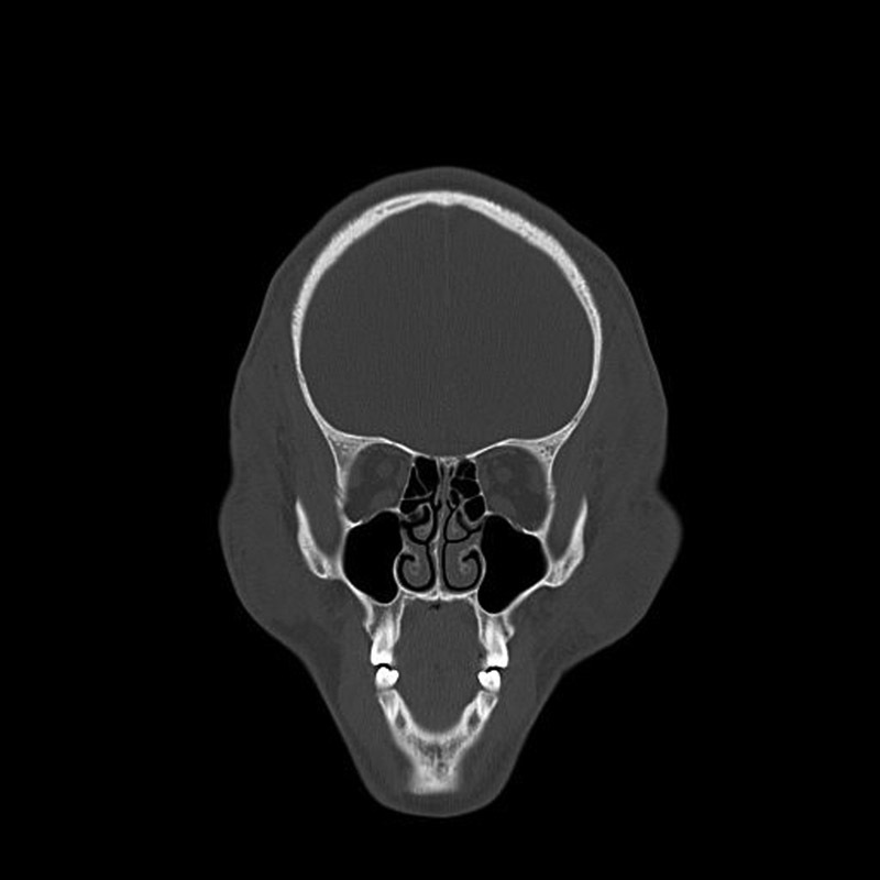
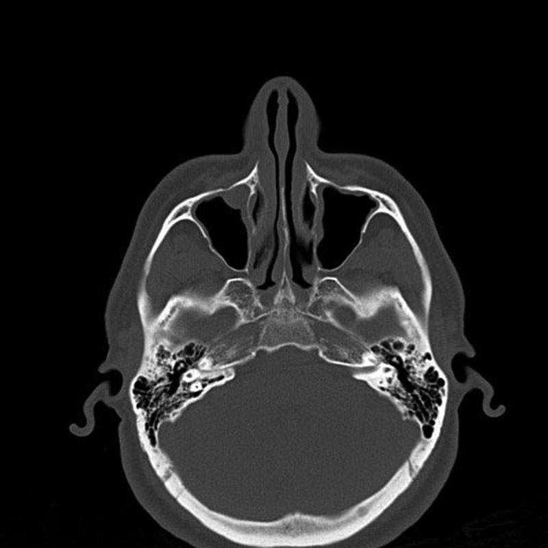
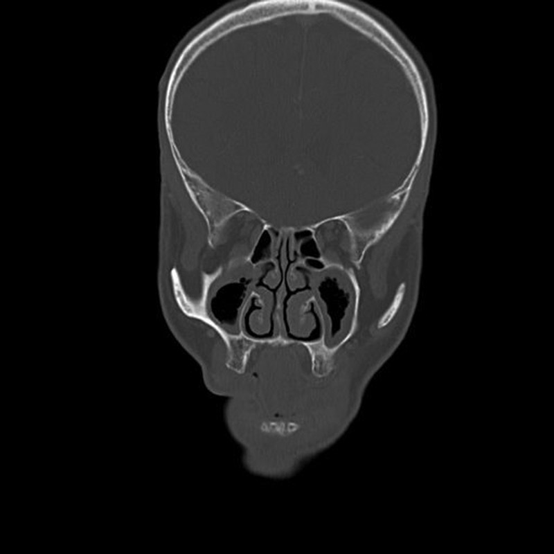
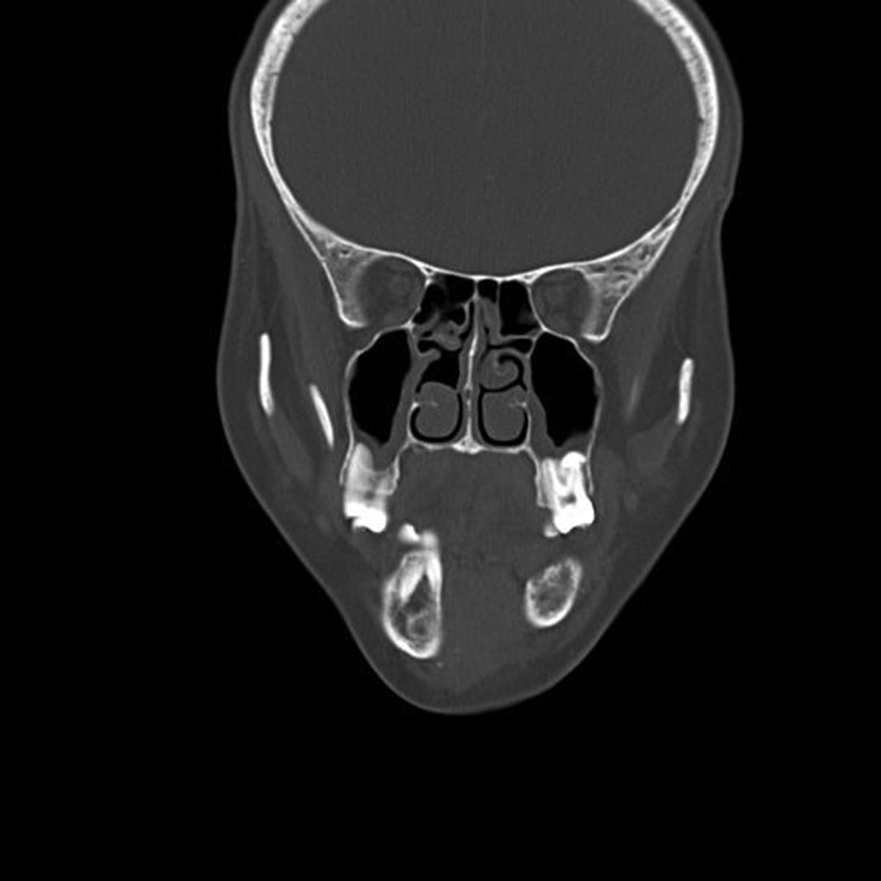
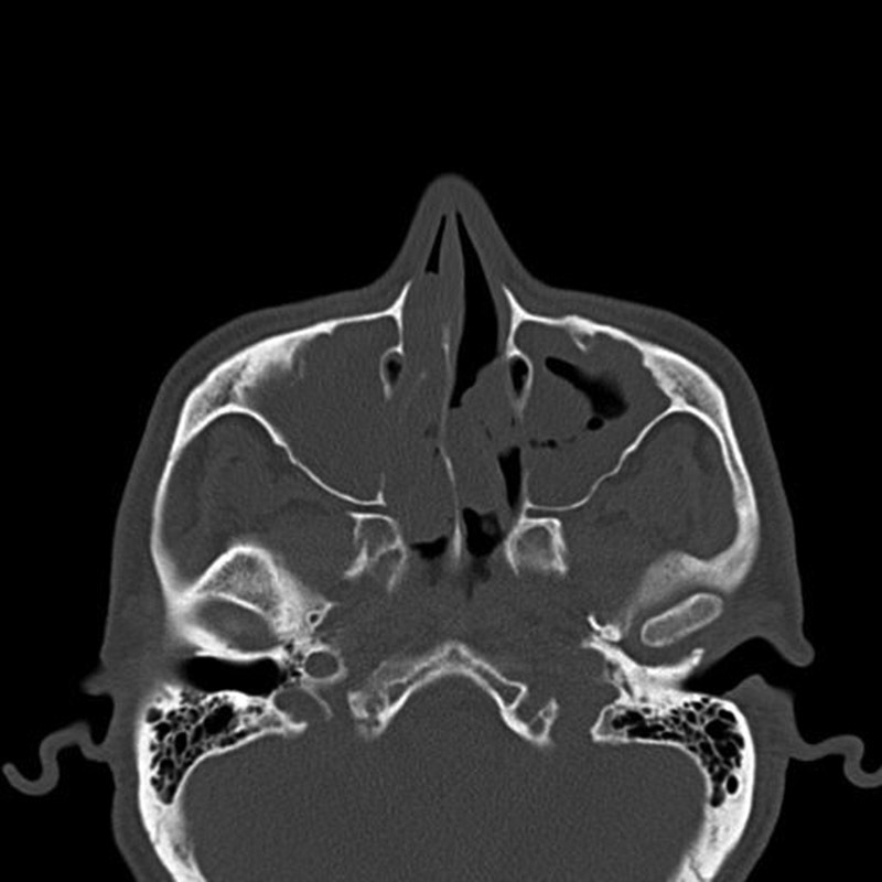
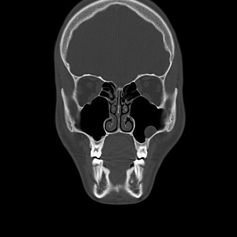
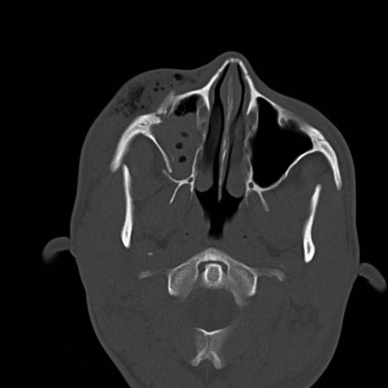
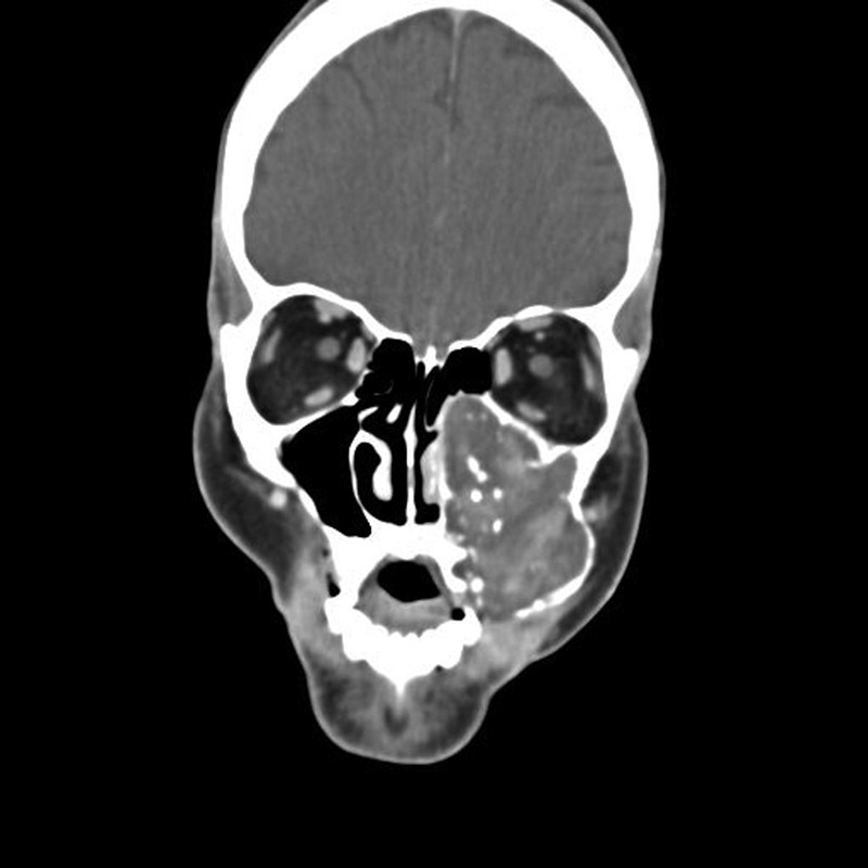
Introduction Maxillary sinus disease is common and numerous disorders can affect this anatomical area. Abnormalities can be classified as: non-neoplastic, neoplastic benign, and neoplastic malignant. Objective Evaluate through CT the prevalence of diseases in maxillary sinuses, using the Radiology Department's database of a hospital in São Paulo city. Methods The sample consisted of 762 facial CT scans that we divided into three groups: Group A (12-19 years old); Group B (20-49 years old); Group C (above 50 years old); and male or female. We considered the following pathological processes: I - Mucoperiosteal Thickening; II - Chronic Sinusitis; III - Chronic Odontogenic Sinusitis; IV - Rhinosinusitis; V - Polypoid Lesions; VI - Bone Lesions; VII - Neoplasms; VIII - Antrolith; IX - Foreign Bodies; X - Oroantral Fistula. Results Our study found that 305 exams (40.02%) were normal and 457 exams (59.97%) were abnormal. We found the following disease frequencies: focal mucoperiosteal thickening (21.25%); polypoid lesions (10.76%); chronic sinusitis (7.48%); chronic odontogenic sinusitis (2.29%); neoplasms (2.03%); rhinosinusitis (1.77%); bone lesions, foreign bodies and oroantral fistula in 0.65%; 0.13% and 0.06% respectively. There was no significant difference between male and female, and Groups A, B, or C when relating the frequencies of abnormalities found. There was no significant difference between male and female and the age group for the side of the altered maxillary sinus. Conclusion We observed a high prevalence of sinus maxillary diseases. Mucoperiosteal thickening; acute, chronic, and odontogenic sinusitis; polypoid lesions and neoplasms have high prevalence in maxillary sinuses. Thus, facial CT exam was effective for the evaluation of diseases in maxillary sinuses.
Keywords: diagnostic imaging; epidemiology; maxillary sinus; otolaryngology; spiral computed tomography.
Figures
Similar articles
-
[Volumetric measurement of the maxillary sinus by coronal CT scan].Nihon Jibiinkoka Gakkai Kaiho. 1996 Aug;99(8):1136-43. doi: 10.3950/jibiinkoka.99.1136. Nihon Jibiinkoka Gakkai Kaiho. 1996. PMID: 8831237 Japanese.
-
Contralateral maxillary sinus lesions in patients with nasal cavity and/or paranasal sinus carcinoma: analysis of computed tomography findings.Ann Otol Rhinol Laryngol. 2008 Dec;117(12):909-13. doi: 10.1177/000348940811701208. Ann Otol Rhinol Laryngol. 2008. PMID: 19140537
-
Frequency of a dental source for acute maxillary sinusitis.Laryngoscope. 2009 Mar;119(3):580-4. doi: 10.1002/lary.20095. Laryngoscope. 2009. PMID: 19160401
-
Imaging of odontogenic sinusitis.Clin Radiol. 2019 Jul;74(7):503-516. doi: 10.1016/j.crad.2019.02.012. Epub 2019 Mar 27. Clin Radiol. 2019. PMID: 30926134 Review.
-
Odontogenic sinusitis: an ancient but under-appreciated cause of maxillary sinusitis.Curr Opin Otolaryngol Head Neck Surg. 2012 Feb;20(1):24-8. doi: 10.1097/MOO.0b013e32834e62ed. Curr Opin Otolaryngol Head Neck Surg. 2012. PMID: 22157162 Review.
Cited by
-
The maxillary sinus: physiology, development and imaging anatomy.Dentomaxillofac Radiol. 2019 Dec;48(8):20190205. doi: 10.1259/dmfr.20190205. Epub 2019 Aug 13. Dentomaxillofac Radiol. 2019. PMID: 31386556 Free PMC article. Review.
-
CBCT Evaluation of Periapical Pathologies in Maxillary Posterior Teeth and Their Relationship with Maxillary Sinus Mucosal Thickening.Healthcare (Basel). 2023 Mar 7;11(6):787. doi: 10.3390/healthcare11060787. Healthcare (Basel). 2023. PMID: 36981444 Free PMC article.
-
A study on the association between accessory maxillary ostium and maxillary sinus mucosal thickening using cone beam computed tomography.Head Face Med. 2021 Jul 14;17(1):28. doi: 10.1186/s13005-021-00284-0. Head Face Med. 2021. PMID: 34261509 Free PMC article.
-
Performance of panoramic radiography compared with computed tomography in the evaluation of pathological changes in the maxillary sinuses: a systematic review and meta-analysis.Dentomaxillofac Radiol. 2023 Jul;52(5):20230067. doi: 10.1259/dmfr.20230067. Epub 2023 May 16. Dentomaxillofac Radiol. 2023. PMID: 37192021 Free PMC article.
-
The Relationship between an Accessory Maxillary Ostium and Variations in Structures Adjacent to the Maxillary Sinus without Polyps.Int Arch Otorhinolaryngol. 2022 Jan 28;26(4):e548-e555. doi: 10.1055/s-0042-1742325. eCollection 2022 Oct. Int Arch Otorhinolaryngol. 2022. PMID: 36405481 Free PMC article.
References
-
- Ogle O E, Weinstock R J, Friedman E.Surgical anatomy of the nasal cavity and paranasal sinuses Oral Maxillofac Surg Clin North Am 20122402155–166., vii - PubMed
-
- Van Dis M L, Miles D A. Disorders of the maxillary sinus. Dent Clin North Am. 1994;38(01):155–166. - PubMed
-
- Stephens J C, Saleh H A. Evaluation and treatment of isolated maxillary sinus disease. Curr Opin Otolaryngol Head Neck Surg. 2013;21(01):50–57. - PubMed
-
- Lawson W, Patel Z M, Lin F Y. The development and pathologic process that influence maxillary sinus pneumatization. Anat Rec. 2008;291:1154–1163. - PubMed
-
- Mehra P, Jeong D. Maxillary sinusitis of odontogenic origin. Curr Allergy Asthma Rep. 2009;9(03):238–243. - PubMed
LinkOut - more resources
Full Text Sources
Other Literature Sources