. 2017 Mar 6:13:417-427.
doi: 10.3762/bjoc.13.45. eCollection 2017.

Structure-efficiency relationships of cyclodextrin scavengers in the hydrolytic degradation of organophosphorus compounds

Affiliations

Structure-efficiency relationships of cyclodextrin scavengers in the hydrolytic degradation of organophosphorus compounds

Sophie Letort et al. Beilstein J Org Chem. .

Abstract

New derivatives of cyclodextrins were prepared in order to determine the relative importance of the structural key elements involved in the degradation of organophosphorus nerve agents. To avoid a competitive inclusion between the organophosphorus substrate and the iodosobenzoate group, responsible for its degradation, the latter group had to be covalently bound to the cyclodextrin scaffold. Although the presence of the α nucleophile iodosobenzoate was a determinant in the hydrolysis process, an imidazole group was added to get a synergistic effect towards the degradation of the agents. The degradation efficiency was found to be dependent on the relative position of the heterocycle towards the reactive group as well as on the nature of the organophosphorus derivative.

Keywords: cyclodextrin; decontamination; enzyme mimic; nerve agents; organophosphorus pesticides.


Figures

Figure 1
Figure 1
Structures of G agents.
Figure 2
Figure 2
Scavenger based on a heterodifunctionalized β-cyclodextrin derivative.
Figure 3
Figure 3
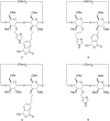
Structures of β-cyclodextrin derivatives 25.
Figure 4
Figure 4
Structures of pesticides tested.
Scheme 1
Scheme 1
Synthetic pathway to derivatives 2 and 3 (Tr = trityl).
Scheme 2
Scheme 2
Synthesis of compound 4.
Scheme 3
Scheme 3
Synthesis of compound 5 (Tr = trityl).
Figure 5
Figure 5
Hydrolysis of methyl paraoxon (0.5 mM) in the presence of compounds 1, 2, 3 or 2-iodosobenzoic acid (IBA) at 0.25 mM concentration.
Figure 6
Figure 6
Hydrolysis of methyl paraoxon (0.5 mM) in the presence of compounds 1, 2, 3 or 2-iodosobenzoic acid (IBA) at 0.5 mM concentration.
Figure 7
Figure 7
Hydrolysis of methyl paraoxon (0.5 mM) in the presence of compounds 2, 4, 5 or 2-iodosobenzoic acid (IBA) at 0.5 mM concentration.
Figure 8
Figure 8
Hydrolysis of methyl paraoxon (0.5 mM) in the presence of mixtures of compounds 4, 5 with IBA or imidazole and in the presence of a mixture of TRIMEB, 2-iodosobenzoic acid (IBA) and imidazole. The final concentrations of compounds 4, 5, 2-iodosobenzoic acid (IBA), imidazole and TRIMEB were 0.5 mM.
Figure 9
Figure 9
Influence of the pesticide structure on the hydrolytic efficiency of compound 2 (0.25 mM). Kinetic assays were carried out with methyl paraoxon, methyl parathion or fenitrothion (0.5 mM).
Figure 10
Figure 10
Influence of TRIMEB, IBA and imidazole on the hydrolysis of methyl parathion (0.5 mM). The final concentrations of compounds 2, 4, 2-iodosobenzoic acid (IBA), imidazole and TRIMEB were 0.25 mM.
Figure 11
Figure 11
Ability of compounds 14 in preventing the inhibition of acetylcholinesterase by soman (GD).

References

    1. Worek F, Thiermann H, Szinicz L, Eyer P. Biochem Pharmacol. 2004;68:2237–2248. doi: 10.1016/j.bcp.2004.07.038. - DOI - PubMed
    1. Tuovinen K. Toxicology. 2004;196:31–39. doi: 10.1016/j.tox.2003.10.013. - DOI - PubMed
    1. Benschop H P, De Jong L P A. Acc Chem Res. 1988;21:368–374. doi: 10.1021/ar00154a003. - DOI
    1. Aragay G, Pino F, Merkoçi A. Chem Rev. 2012;112:5317–5338. doi: 10.1021/cr300020c. - DOI - PubMed
    1. Taylor P, Radic Z, Hosea N A, Camp S, Marchot P, Berman H A. Toxicol Lett. 1995;82–83:453–458. doi: 10.1016/0378-4274(95)03575-3. - DOI - PubMed

LinkOut - more resources