

Sonographic Evaluation of Structural Changes in Post-Stroke Hemiplegic Shoulders
- PMID: 28382186
- PMCID: PMC5360429
- DOI: 10.12659/PJR.899684
Sonographic Evaluation of Structural Changes in Post-Stroke Hemiplegic Shoulders
Abstract
Background: Stroke and hemiplegia are frequent complications of stroke. This study was performed to sonographically evaluate post-stroke hemiplegic shoulders and explore possible relationship(s) between the sonographic findings and clinical indices.
Material/methods: Forty-five stroke patients and 45 age- and sex-matched controls were recruited. Standard sonographic examination of both shoulders was performed to assess for joint subluxation, rotator cuff tears, tendinosis, subacromial-subdeltoid bursitis or effusion and adhesive capsulitis.
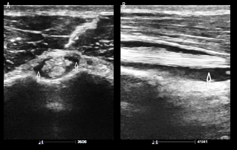
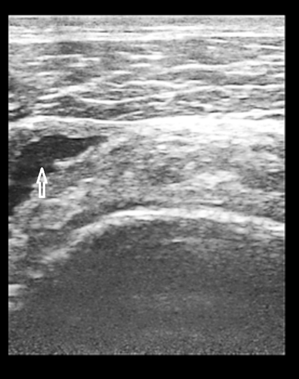
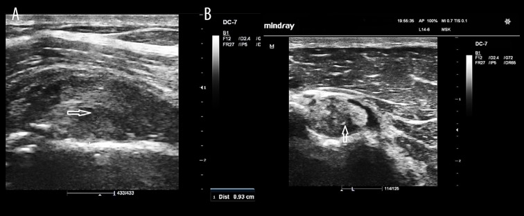
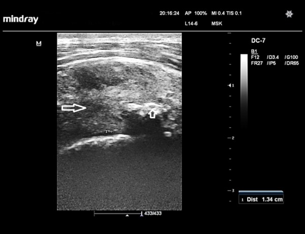
Results: Hemiplegic shoulders exhibited significantly higher number of pathologies compared to the unaffected shoulders and shoulders of controls (p=0.000). One or more structural abnormalities were found in all 45 (100%) hemiplegic shoulders, 25 (55.6%) unaffected shoulders of the stroke subjects, and 39 (43.3%) control shoulders. The most frequent pathologies in the hemiplegic shoulders were the following: tendinosis of the long head of bicep tendon (48.9%), inferior shoulder subluxation (44.4%), co-existing subacromial-subdeltoid bursa/long head of bicep tendon sheath effusion (44.4%), and long head of bicep tendon sheath effusion only (40%). Tendinosis of the long head of bicep tendon was commoner in hemiplegic shoulders with poor motor status than those with good motor status.
Conclusions: Hemiplegic shoulders have significantly higher number of structural abnormalities than unaffected shoulders and the shoulders of controls. Hemiplegic stroke patients should undergo ultrasonography of the hemiplegic shoulder to define the nature and extent of soft tissue injuries prior to physical therapy.
Keywords: Hemiplegia; Shoulder Joint; Stroke; Ultrasonography.
Figures
References
-
- Lindgren I, Jönsson AC, Norrving B, Lindgren A. Shoulder pain after stroke: A prospective population-based study. Stroke. 2007;38:343–48. - PubMed
-
- Lee IS, Shin YB, Moon T, et al. Sonography of patients with hemiplegic shoulder pain after stroke: correlation with motor recovery stage. Am J Roentgenol. 2009;192:W40–44. - PubMed
-
- De Jesus JO, Parker L, Frangos AJ, Nazarian LN. Accuracy of MRI, MR arthrography, and ultrasound in the diagnosis of rotator cuff tears: A meta-analysis. Am J Roentgenol. 2009;192:1701–7. - PubMed
-
- Lazarian LN. The top 10 reasons musculoskeletal sonography is an important complementary or alternative technique to MRI. Am J Roentgenol. 2008;190:1621–26. - PubMed
-
- Middleton WD, Payne WT, Teefey SA, et al. Sonography and MRI of the shoulder: Comparison of patient satisfaction. Am J Roentgenol. 2004;183:1449–52. - PubMed
LinkOut - more resources
Full Text Sources
Other Literature Sources
Medical