Review
. 2017 Sep;469(9):1039-1050.
doi: 10.1007/s00424-017-1968-0. Epub 2017 Apr 5.

Biology of pancreatic stellate cells-more than just pancreatic cancer

Affiliations
Review

Biology of pancreatic stellate cells-more than just pancreatic cancer

Pawel E Ferdek et al. Pflugers Arch. 2017 Sep.

Abstract

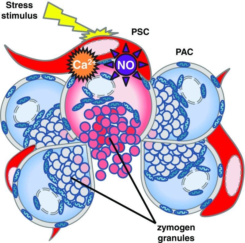
Pancreatic stellate cells, normally quiescent, are capable of remarkable transition into their activated myofibroblast-like phenotype. It is now commonly accepted that these cells play a pivotal role in the desmoplastic reaction present in severe pancreatic disorders. In recent years, enormous scientific effort has been devoted to understanding their roles in pancreatic cancer, which continues to remain one of the most deadly diseases. Therefore, it is not surprising that considerably less attention has been given to studying physiological functions of pancreatic stellate cells. Here, we review recent advances not only in the field of pancreatic stellate cell pathophysiology but also emphasise their roles in physiological processes.

Keywords: Calcium; Fibrosis; Myofibroblasts; Pancreatic cancer; Pancreatic stellate cells; Pancreatitis.


Conflict of interest statement

The authors declare that they have no conflict of interest.

Figures

Fig. 1
Fig. 1
Schematic illustration of the pancreas. The exocrine part of the organ predominantly consists of acinar lobules. Pancreatic stellate cells (shown in bright green-yellow-red pseudocolours, lower panel) form a three-dimensional network in between those lobules (dark purple)
Fig. 2
Fig. 2
Pancreatic stellate cells have the capacity to store retinoids. Cultured human PSCs quickly become activated and lose most of their stored retinol (upper panel). In the presence of 100 μM retinol (24 h treatment), lipid vesicles appear in the cytosol of PSCs (lower panel, red pattern), which is revealed by excitation with UV light
Fig. 3
Fig. 3
Expression of α-smooth muscle actin (α-SMA), and thus the number of activated PSCs (aPSCs), increases as a result of tissue inflammation. In the healthy mouse pancreas (upper panel) α-SMA-positive staining (red) is only present in the vascular smooth muscle cells in the blood vessels and is labelled as vascular smooth muscle actin (VSMA). Induction of pancreatitis (by ethanol and fatty acids) leads to a sudden appearance of α-SMA-positive cells—aPSCs—scattered within the parenchymal tissue
Fig. 4
Fig. 4
Mouse pancreatic stellate cells, in their native environment of pancreatic lobules, respond to pathophysiological stimuli with intracellular Ca2+ signals as well as generation of NO. a Sample traces recorded in mouse pancreatic lobules loaded with a Ca2+-sensitive dye Fluo-4 AM. Pancreatic stellate cell (PSC, red trace) responds to 10 nM bradykinin (BK) but pancreatic acinar cell (PAC, blue trace) does not, which confirms the stellate phenotype. The PSC subsequently responds to 5 mM taurocholate (TC) with a large elevation of intracellular Ca2+, whereas the neighbouring PAC generates only modest Ca2+ oscillations. For more information, the reader is referred to a study by Ferdek et al. [30]. b Individual images from the recording shown in (a). The red circular regions mark the PSC that responded to bradykinin and then to taurocholate with increases in intracellular Ca2+ concentration. The blue circular regions indicate the PAC that did not respond to bradykinin and produced only transient Ca2+ elevations in response to treatment with taurocholate. c Sample traces recorded in a PSC embedded in a mouse pancreatic lobule loaded with both Fura-2 AM (Ca2+-sensitive dye) and DAF-2 (NO-sensitive dye). The cell responds to 20 nM BK with an elevation of intracellular Ca2+ concentration (red trace) and a simultaneous increase in intracellular NO (purple trace). For more information, the reader is referred to a study by Jakubowska et al. [58]. d Sample images show a mouse pancreatic lobule, loaded with DAF-2, before and after treatment with 500 μM hydrogen peroxide (H2O2). PSCs are indicated with white arrowheads. Treatment with H2O2 increases intracellular NO in these cells (shown as a shift in the pseudocolour spectrum)
Fig. 5
Fig. 5
Schematic illustration of a pancreatic lobule. Pathophysiological stimuli (e.g. bile acids, bradykinin, H2O2) induce stress responses in pancreatic stellate cells (PSC, red), manifested as an increase in the cytosolic Ca2+ concentration and NO generation. Stress in PSCs further escalates pathophysiological responses in adjacent pancreatic acinar cells (PAC, blue) leading to premature activation of zymogen granules, exacerbated inflammation and necrotic cell death (pink), associated with loss of the cellular content

References

    1. Andoh A, Takaya H, Saotome T, Shimada M, Hata K, Araki Y, Nakamura F, Shintani Y, Fujiyama Y, Bamba T. Cytokine regulation of chemokine (IL-8, MCP-1, and RANTES) gene expression in human pancreatic periacinar myofibroblasts. Gastroenterology. 2000;119:211–219. doi: 10.1053/gast.2000.8538. - DOI - PubMed
    1. Apte MV, Haber PS, Applegate TL, Norton ID, McCaughan GW, Korsten MA, Pirola RC, Wilson JS. Periacinar stellate shaped cells in rat pancreas: identification, isolation, and culture. Gut. 1998;43:128–133. doi: 10.1136/gut.43.1.128. - DOI - PMC - PubMed
    1. Apte MV, Haber PS, Darby SJ, Rodgers SC, McCaughan GW, Korsten MA, Pirola RC, Wilson JS. Pancreatic stellate cells are activated by proinflammatory cytokines: implications for pancreatic fibrogenesis. Gut. 1999;44:534–541. doi: 10.1136/gut.44.4.534. - DOI - PMC - PubMed
    1. Apte MV, Phillips PA, Fahmy RG, Darby SJ, Rodgers SC, McCaughan GW, Korsten MA, Pirola RC, Naidoo D, Wilson JS. Does alcohol directly stimulate pancreatic fibrogenesis? Studies with rat pancreatic stellate cells. Gastroenterology. 2000;118:780–794. doi: 10.1016/S0016-5085(00)70148-X. - DOI - PubMed
    1. Apte MV, Park S, Phillips PA, Santucci N, Goldstein D, Kumar RK, Ramm GA, Buchler M, Friess H, McCarroll JA, Keogh G, Merrett N, Pirola R, Wilson JS. Desmoplastic reaction in pancreatic cancer: role of pancreatic stellate cells. Pancreas. 2004;29:179–187. doi: 10.1097/00006676-200410000-00002. - DOI - PubMed

Publication types

LinkOut - more resources