. 2017 Oct;32(5):404-418.
doi: 10.1111/omi.12182. Epub 2017 May 22.

Behavior of two Tannerella forsythia strains and their cell surface mutants in multispecies oral biofilms

Affiliations

Behavior of two Tannerella forsythia strains and their cell surface mutants in multispecies oral biofilms

Susanne Bloch et al. Mol Oral Microbiol. 2017 Oct.

Abstract

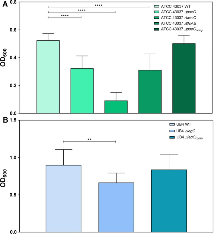
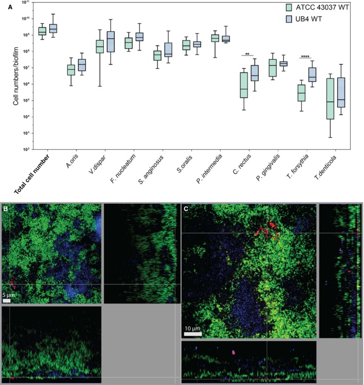
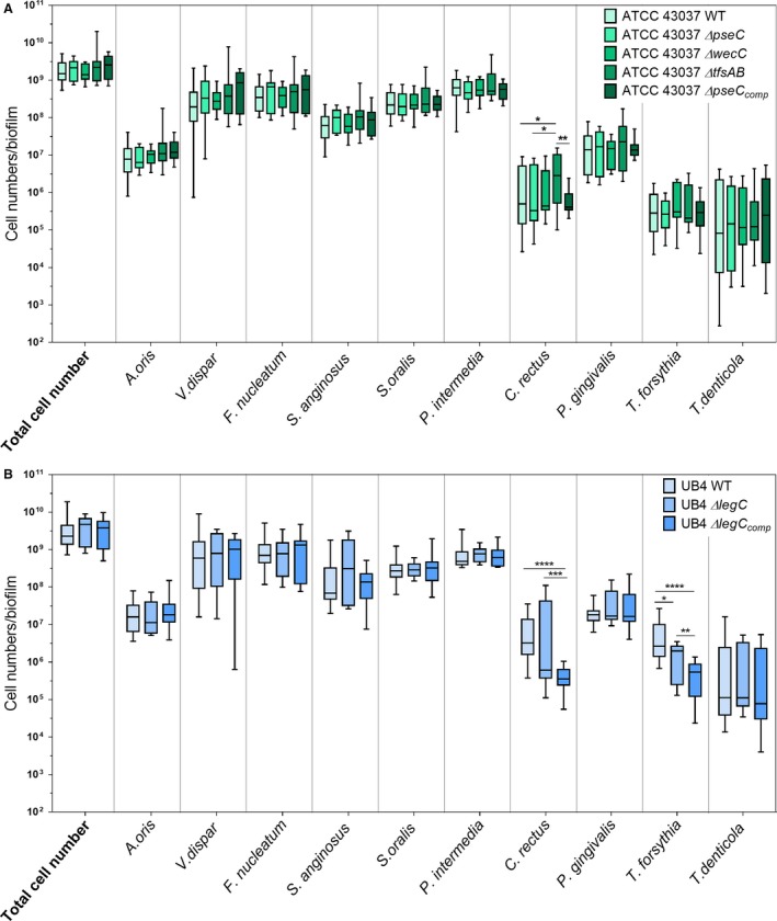
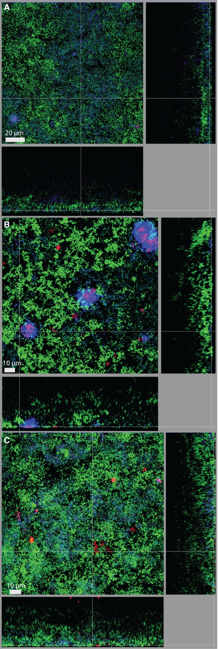
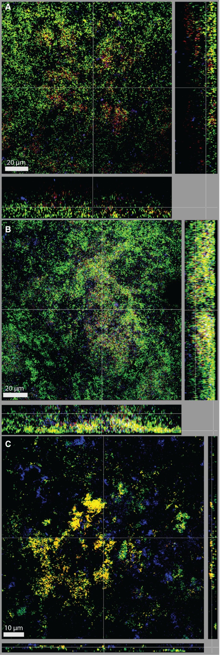
As a member of subgingival multispecies biofilms, Tannerella forsythia is commonly associated with periodontitis. The bacterium has a characteristic cell surface (S-) layer modified with a unique O-glycan. Both the S-layer and the O-glycan were analyzed in this study for their role in biofilm formation by employing an in vitro multispecies biofilm model mimicking the situation in the oral cavity. Different T. forsythia strains and mutants with characterized defects in cell surface composition were incorporated into the model, together with nine species of select oral bacteria. The influence of the T. forsythia S-layer and attached glycan on the bacterial composition of the biofilms was analyzed quantitatively using colony-forming unit counts and quantitative real-time polymerase chain reaction, as well as qualitatively by fluorescence in situ hybridization and confocal laser scanning microscopy. This revealed that changes in the T. forsythia cell surface did not affect the quantitative composition of the multispecies consortium, with the exception of Campylobacter rectus cell numbers. The localization of T. forsythia within the bacterial agglomeration varied depending on changes in the S-layer glycan, and this also affected its aggregation with Porphyromonas gingivalis. This suggests a selective role for the glycosylated T. forsythia S-layer in the positioning of this species within the biofilm, its co-localization with P. gingivalis, and the prevalence of C. rectus. These findings might translate into a potential role of T. forsythia cell surface structures in the virulence of this species when interacting with host tissues and the immune system, from within or beyond the biofilm.

Keywords: Campylobacter rectus; Tannerella forsythia; S-layer glycosylation; cell surface; oral biofilm; periodontal disease.


Figures

Figure 1
Figure 1
Monospecies biofilm formation of Tannerella forsythia wild‐type and mutant strains. (A) Biofilm formation of T. forsythia ATCC 43037 wild‐type compared with its mutants ATCC 43037 ΔpseC, ΔwecC, ΔtfsAB and the complemented mutant ΔpseC comp. (B) Biofilm formation of T. forsythia UB4 wild‐type compared with its mutant UB4 ΔlegC and the complemented mutant ΔlegC comp. Mean values ±SD of four independent experiments with three replicates, each, are shown. Asterisks (**) indicate significant differences between samples as determined by the unpaired Student's t‐test (P≤.01)
Figure 2
Figure 2
Comparison of colony‐forming unit (CFU) counting and quantitative polymerase chain reaction (qPCR) for Tannerella forsythia wild‐type strains and mutants in the subgingival “Zurich biofilm”. Total bacteria for 10‐species biofilms with different T. forsythia strains and mutants enumerated by CFU counts (red boxes) and qPCR (blue boxes) for three independent experiments with three technical replicates, each, are shown (Whiskers boxplots 5th to 95th centile)
Figure 3
Figure 3
Comparison of 10‐species biofilms with two Tannerella forsythia wild‐type strains. (A) Whiskers boxplots (5th to 95th centile) show bacterial numbers determined by quantitative real‐time PCR from three independent experiments. Asterisk (*) indicates a statistically significant difference (P≤.05) between groups. The two groups represent biofilms with either T. forsythia ATCC 43037 wild‐type or T. forsythia UB4 wild‐type. (B, C) Fluorescence in situ hybridization stainings of fixed biofilms showing the localization of ATCC 43037 wild‐type (B) and UB4 wild‐type (C). Red/yellow: T. forsythia; cyan: Porphyromonas gingivalis, green: non‐hybridized cells (DNA staining YoPro‐1+Sytox). Here a representative area for one disk each is shown with a top view in the left panel and a side view with the biofilm–disk interface directed towards the top view; scale bars 5 μm (B) and 10 μm (C)
Figure 4
Figure 4
Box plots showing cell numbers of all species determined by quantitative real‐time PCR for biofilms with Tannerella forsythia ATCC 43037 wild‐type or mutants (∆pseC, ∆wecC, ∆tfsAB, ∆pseC comp) (A) and UB4 wild‐type or mutants (∆legC, ∆legC comp), respectively (B). Data derived from three independent experiments were plotted on a logarithmic scale. Asterisk (*) indicates significant differences (P≤.05) between the groups
Figure 5
Figure 5
Fluorescence in situ hybridization staining of biofilms harboring Tannerella forsythia ATCC 43037 mutants (A) ∆pseC, (B) ∆wecC, and (C) ∆tfsAB. Red: T. forsythia, cyan: Porphyromonas gingivalis, green: non‐hybridized cells (DNA staining YoPro‐1+Sytox). Scale bars 20 μm (A) and 10 μm (B, C)
Figure 6
Figure 6
Dual fluorescence in situ hybridization staining of Tannerella forsythia and Campylobacter rectus for biofilms harboring ATCC 43037 wild‐type (A), UB4 wild‐type (B), and ATCC 43037 ∆tfsAB (C). Red/yellow: T. forsythia, cyan: C. rectus; green: non‐hybridized cells (DNA staining YoPro‐1+Sytox). Scale bars 20 μm (A, B) and 15 μm (C)

References

    1. Marsh PD. Dental plaque: Biological significance of a biofilm and community life‐style. J Clin Periodontol. 2005;32:7‐15. - PubMed
    1. Marsh PD. Dental plaque as a microbial biofilm. Caries Res. 2004;38:204‐211. - PubMed
    1. Van Dyke TE, Sheilesh D. Risk factors for periodontitis. J Int Acad Periodontol. 2005;7:3‐7. - PMC - PubMed
    1. Hajishengallis G, Lamont RJ. Beyond the red complex and into more complexity: The polymicrobial synergy and dysbiosis (PSD) model of periodontal disease etiology. Mol Oral Microbiol. 2012;27:409‐419. - PMC - PubMed
    1. Socransky SS, Haffajee AD, Cugini MA, Smith C, Kent RL Jr. Microbial complexes in subgingival plaque. J Clin Periodontol. 1998;25:134‐144. - PubMed

MeSH terms

LinkOut - more resources