. 2017 Apr 6;8(4):e2729.
doi: 10.1038/cddis.2017.153.

Extracellular cystatin SN and cathepsin B prevent cellular senescence by inhibiting abnormal glycogen accumulation

Affiliations

Extracellular cystatin SN and cathepsin B prevent cellular senescence by inhibiting abnormal glycogen accumulation

Sang-Seok Oh et al. Cell Death Dis. .

Abstract

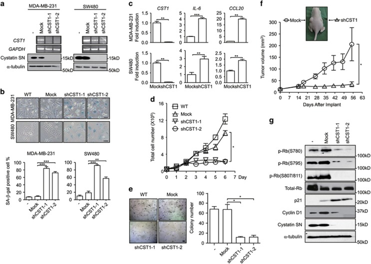
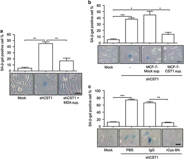
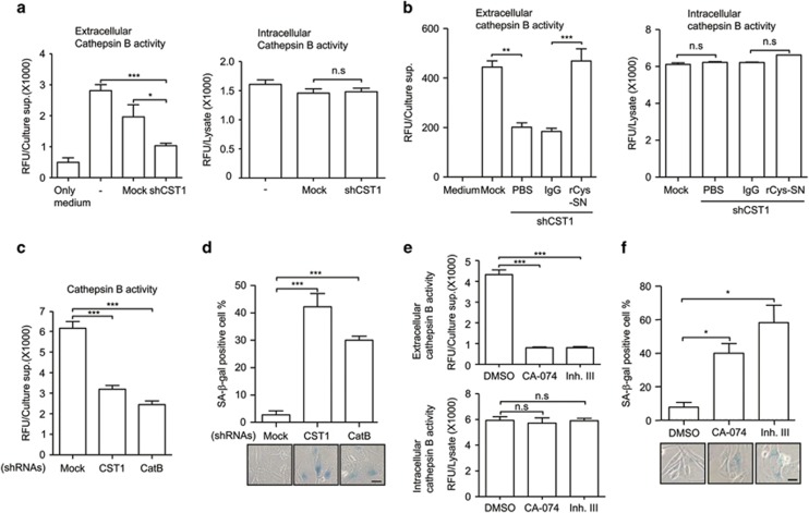
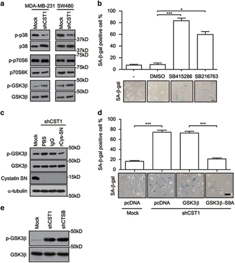
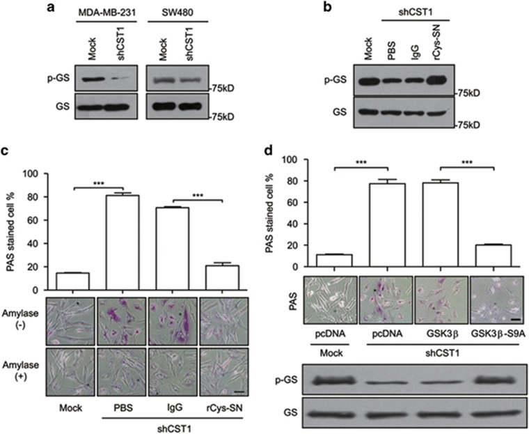
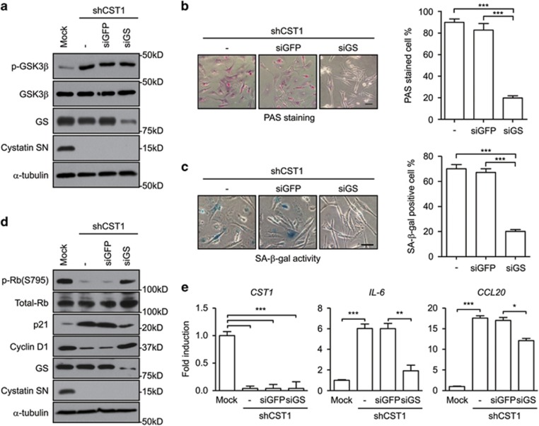
Cystatin SN (CST1), a known inhibitor of cathepsin B (CatB), has important roles in tumor development. Paradoxically, CatB is a member of the cysteine cathepsin family that acts in cellular processes, such as tumor development and invasion. However, the relationship between CST1 and CatB, and their roles in tumor development are poorly understood. In this study, we observed that the knockdown of CST1 induced the activity of senescence-associated β-galactosidase, a marker of cellular senescence, and expression of senescence-associated secretory phenotype genes, including interleukin-6 and chemokine (C-C motif) ligand 20, in MDA-MB-231 and SW480 cancer cells. Furthermore, CST1 knockdown decreased extracellular CatB activity, and direct CatB inhibition, using specific inhibitors or shCatB, induced cellular senescence. Reconstitution of CST1 restored CatB activity and inhibited cellular senescence in CST1 knockdown cells. CST1 knockdown or CatB inhibition increased glycogen synthase (GS) kinase 3β phosphorylation at serine 9, resulting in the activation of GS and the induction of glycogen accumulation associated with cellular senescence. Importantly, CST1 knockdown suppressed cancer cell proliferation, soft agar colony growth and tumor growth in a xenograft model. These results indicate that CST1-mediated extracellular CatB activity enhances tumor development by preventing cellular senescence. Our findings suggest that antagonists of CST1 or inhibitors of CatB are potential anticancer agents.


Conflict of interest statement

The authors declare no conflict of interest.

Figures

Figure 1
Figure 1
CST1 is required for cell proliferation and colony growth in vitro and in vivo. The following assays were performed in human cancer MDA-MB-231 and SW480 cells transduced with a control shRNA (mock) or an shRNA for CST1. (a) The efficiency of CST1 knockdown was confirmed by reverse transcription PCR (upper) and western blotting (lower). (b) Representative photographs of SA-β-gal staining (upper). Senescence under the indicated condition was quantified as the percentage of SA-β-gal-positive cells (lower). Scale bars, 50 μm. (c) The expression of SASP genes induced by CST1 knockdown. RNAs were extracted from cells, and real-time PCR analysis was performed to quantify the transcripts of CST1, IL-6 and chemokine ligand 20 (CCL20). (d) Analysis of anchorage-dependent cell proliferation. Cells were seeded on to six-well plates (5 × 10⁴ cells per well) and cultured for the indicated number of days. The number of cells was counted using a hemocytometer. One-way ANOVA was used for statistical analysis (*P<0.05). (e) Analysis of anchorage-independent cell proliferation. Cells were seeded on to complete DMEM containing agarose (1 × 10⁵ cells per well) and cultured for 12 days. Formed colonies were stained with 0.005% crystal violet and quantified under a light microscope. Scale bar, 10 μm. (f) Analysis of tumor growth in the xenograft model. MDA-MB-231 cells transduced with a control shRNA or shCST1 (1 × 10⁶ cells) were subcutaneously implanted into the left or right flanks of athymic nude mice (n=8). Tumor volume was measured on the indicated days. One-way ANOVA was used for statistical analysis (*P<0.05). (g) Protein expression of p-Rb, total Rb, p21, cyclin D1 or CST1 was evaluated by western blotting. *P<0.05; **P<0.01; ***P<0.001 by Student's t-test
Figure 2
Figure 2
Extracellular CST1 is required in the prevention of cellular senescence. MDA-MB-231 cells transduced with a control shRNA (mock) or an shRNA for CST1 were cultured with an MDA-MB-231 culture supernatant (a) or CST1-overexpressing MCF-7 culture supernatant (b). (c) The effect of recombinant CST1-immunoglobulin (rCys-SN) on CST1 knockdown-mediated cellular senescence. Scale bars, 50 μm. *P<0.05; **P<0.01; ***P<0.001 by Student's t-test
Figure 3
Figure 3
Downregulation of CatB activity by CST1 knockdown induces cellular senescence. (a) CatB activity was measured in the culture supernatant (left) or cell lysate (right) of CST1 knockdown cells. CatB activity of the culture supernatant (left) or cell lysate (right) from rCys-SN treated cells (b), or culture supernatant of CatB knockdown cells (c) was analyzed using a CatB activity fluorometric assay kit. (d) Effect of CatB knockdown on cellular senescence. After CST1 or CatB knockdown, cellular senescence was quantified by calculating the percentage of SA-β-gal-positive cells. To confirm the function of CatB associated with cellular senescence, MDA-MB-231 cells were treated with CatB inhibitors. The efficacy of the inhibitors was confirmed by the analysis of extracellular or intracellular CatB activity (e), and cellular senescence induced by the inhibitors was quantified by SA-β-gal staining (f). Scale bars, 50 μm. Data were obtained from three independent experiments, and values are presented as the means±S.E.M. *P<0.05; **P<0.01; ***P<0.001 by Student's t-test
Figure 4
Figure 4
CST1 knockdown-mediated cellular senescence is mediated through GSK3β activity. (a) The phosphorylation of each kinase in CST1 knockdown cells was confirmed by western blotting. (b) The effect of GSK3β inhibitors on cellular senescence. MDA-MB-231 cells were cultured for 96 h in the presence of dimethyl sulfoxide or two different GSK3β inhibitors. Cellular senescence was quantified as the percentage of SA-β-gal-positive cells (upper) and photographed (lower). (c) Modulation of inhibitory phospho-GSK3β mediated by extracellular CST1. CST1 knockdown cells were cultured in the absence or presence of recombinant CST1-immunoglobulin (rCys-SN). (d) The inhibitory effect of active GSK3β on CST1 knockdown-mediated cellular senescence. MDA-MB-231 cells were pretransfected with pcDNA-HA, pcDNA-GSK3β-HA, and pcDNA-GSK3β-S9A-HA plasmids 24 h before the transduction of a control shRNA or an shRNA for CST1. (e) Modulation of phopho-GSK3β in CatB knockdown cells. Scale bars, 50 μm. *P<0.05; ***P<0.001 by Student's t-test
Figure 5
Figure 5
Downregulation of GSK3β activity mediated by CST1 knockdown induces cellular glycogen accumulation. (a and b) Modulation of phospho-GS in CST1 knockdown cells. MDA-MB-231 cells were transduced with a control short hairpin (shRNA) or shCST1 (a) and, in some experiments, cells were replenished with rCys-SN (b). (c and d) Analysis of glycogen accumulation in the cytosol. MDA-MB-231 cells were transduced with a control shRNA or shCST1 in the presence or absence of rCys-SN (c) and, in some experiments, transduction was performed after transfection with pcDNA, pcDNA-GSK3β or pcDNA-GSK3β-S9A (d) Glycogen accumulation was quantified as the percentage of PAS staining-positive cells (magenta) and photographed. Scale bars, 50 μm. ***P<0.001 by Student's t-test
Figure 6
Figure 6
Cellular senescence is induced by GS activation resulted in glycogen accumulation in CST1 knockdown cells. The following assays were performed in CST1 knockdown MDA-MB-231 cells transfected with short-interfering RNAs (siRNAs) for green fluorescent protein (GFP) or GS. (a and d) The expression level of the indicated protein was analyzed by western blotting. (b) PAS staining analysis was performed to assess glycogen accumulation. (c) Quantification of cellular senescence was determined by the SA-β-gal assay. (e) Expression of SASP genes, IL-6 and CCL20, was analyzed by real-time polymerase chain reaction. The results were normalized to the level of β-actin. Scale bars, 50 μm. *P<0.05; **, P<0.01, *** P<0.001 by Student's t-test

References

    1. Turk V, Turk B, Guncar G, Turk D, Kos J. Lysosomal cathepsins: structure, role in antigen processing and presentation, and cancer. Adv Enzyme Regul 2002; 42: 285–303. - PubMed
    1. Buck MR, Karustis DG, Day NA, Honn KV, Sloane BF. Degradation of extracellular-matrix proteins by human cathepsin B from normal and tumour tissues. Biochem J 1992; 282: 273–278. - PMC - PubMed
    1. Premzl A, Zavasnik-Bergant V, Turk V, Kos J. Intracellular and extracellular cathepsin B facilitate invasion of MCF-10 A neoT cells through reconstituted extracellular matrix in vitro. Exp Cell Res 2003; 283: 206–214. - PubMed
    1. Roshy S, Sloane BF, Moin K. Pericellular cathepsin B and malignant progression. Cancer Metastasis Rev 2003; 22: 271–286. - PubMed
    1. Bao W, Fan Q, Luo X, Cheng WW, Wang YD, Li ZN et al. Silencing of cathepsin B suppresses the proliferation and invasion of endometrial cancer. Oncol Rep 2013; 30: 723–730. - PubMed

Publication types

MeSH terms

LinkOut - more resources