. 2017 Apr 6;8(4):e86.
doi: 10.1038/ctg.2017.3.

Diversity of Precursor Lesions For Pancreatic Cancer: The Genetics and Biology of Intraductal Papillary Mucinous Neoplasm

Affiliations

Diversity of Precursor Lesions For Pancreatic Cancer: The Genetics and Biology of Intraductal Papillary Mucinous Neoplasm

Krushna C Patra et al. Clin Transl Gastroenterol. .

Abstract

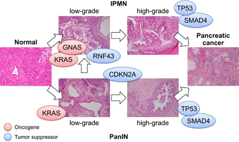
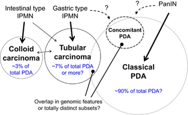
Pancreatic ductal adenocarcinoma (PDA), one of the most lethal cancers worldwide, is associated with two main types of morphologically distinct precursors-pancreatic intraepithelial neoplasia (PanIN) and intraductal papillary mucinous neoplasm (IPMN). Although the progression of PanIN into invasive cancer has been well characterized, there remains an urgent need to understand the biology of IPMNs, which are larger radiographically detectable cystic tumors. IPMNs comprise a number of subtypes with heterogeneous histopathologic and clinical features. Although frequently remaining benign, a significant proportion exhibits malignant progression. Unfortunately, there are presently no accurate prognosticators for assessing cancer risk in individuals with IPMN. Moreover, the fundamental mechanisms differentiating PanIN and IPMN remain largely obscure, as do those that distinguish IPMN subtypes. Recent studies, however, have identified distinct genetic profiles between PanIN and IPMN, providing a framework to better understand the diversity of the precursors for PDA. Here, we review the clinical, biological, and genetic properties of IPMN and discuss various models for progression of these tumors to invasive PDA.


Conflict of interest statement

Guarantor of the article: Yusuke Mizukami, MD, PhD.

Specific author contributions: KCP, NB and YM wrote and edited the manuscript. All the authors have critically reviewed the manuscript. The final version of the manuscript was approved by all the authors.

Financial support: This work was supported by JSPS KAKENHI Grant Number 25461029 and by a Pancreas Research Foundation in Japan to YM and support from the Granara-Skerry Trust, the Linda J. Verville Foundation, the Begg Family, and grants from the NIH (P01 CA117969-07, R01 CA133557-05) to NB. KCP is supported by a post-doctoral fellowship from the Department of Defense, USA (W81XWH-16-1-0285). The authors are Andrew L. Warshaw Institute for Pancreatic Cancer Research scholars. N.B. is the holder of the Gallagher Chair in Gastrointestinal Cancer Research at the Massachusetts General Hospital, USA.

Potential competing interests: None.

Figures

Figure 1
Figure 1
Precursors of pancreatic cancer. Pancreatic ductal adenocarcinoma (PDA) can arise from the progression of IPMN (top) or PanIN (bottom). It is not known whether early IPMNs (incipient IPMN) originate from low-grade PanIN, or develop independently from normal pancreatic ducts or other pancreatic cell lineages. Red and blue circles indicate oncogenes and tumor-suppressor genes, respectively. The precise timing of RNF43 mutations and the order of GNAS and KRAS mutations have not been fully established. IPMN, intraductal papillary mucinous neoplasm; PanIN, pancreatic intraepithelial neoplasia.
Figure 2
Figure 2
Signaling pathways controlling IPMN and associated PDA. (a) Mutant GNAS (encoding the Gαs protein) constitutively activates Adenylyl cyclase (AC), leading to generation of cyclic AMP (cAMP) and consequent induction of diverse intracellular signaling pathways. Effectors of cAMP include protein kinase A (PKA), exchange protein directly activated by cAMP (EPAC1 and EPAC2) and cyclic nucleotide-gated (CNG) channels. The relevant pathways mediating the oncogenic functions of mutant GNAS in pancreatic tumorigenesis have not been elucidated. (b) RNF43 is a transmembrane E3 ubiquitin ligase that inhibits Wnt/β-catenin signaling by reducing the level of Frizzled receptors (Fzd). RNF43 has also been suggested to localize to the nuclear envelope localization where it inactivates Tcf4, a transcription factor that cooperates with β-catenin to induce Wnt target genes. Therefore, loss-of-function mutations in RNF43 can enhance Wnt signaling. Gαs, G-protein alpha stimulatory subunit; IPMN, intraductal papillary mucinous neoplasm; PDA, pancreatic ductal adenocarcinoma.
Figure 3
Figure 3
PDA subtypes and their precursors. Pathologic analysis indicates that intestinal-type IPMN can progress directly to an unusual subtype of invasive pancreatic cancer known as colloid carcinoma. In addition, a subset of, typically gastric- (and pancreatobiliary) type, progress to invasive tubular carcinomas, which are histologically indistinguishable from “classic PDA”. The frequency shown is based on the studies on histologically confirmed IPMN-derived carcinomas and classic PDAs reported in the same time period., , , Moreover, recent studies demonstrated that about 8–11% of PDA harbor GNAS mutations or amplifications, (also see TCGA data set, http://www.cbioportal.org) and IPMN-derived PDA accounts for ~10% of all PDA resected; therefore, ~90% PDA are considered to originate from PanIN, however, there is significant variability between studies. Thus, this estimate largely depends on institutional definition of PanIN and IPMN. Note that the precise origins of concomitant PDA remain to be fully defined (see text for details). IPMN, intraductal papillary mucinous neoplasm; PanIN, pancreatic intraepithelial neoplasia; PDA, pancreatic ductal adenocarcinoma.
Figure 4
Figure 4
Human PanINs observed in resected specimens of PDA. Classic PDAs are often accompanied by multiple PanIN-like lesions, with varying grade and morphology. The presence of these microscopic lesions have been the subject of investigation as potentially predicting post-surgery recurrence. Such lesions may include early IPMNs, as some have been reported to harbor GNAS mutations (see refs 31, 48). There are two patterns of morphology. (a) Solitary PanINs observed in normal pancreatic tissue (acinar compartment without pancreatitis) apart from PDA. (b) PanINs (arrowheads) associated with acinar-to-ductal metaplasia (ADM). This pattern is more frequently seen in IPMN patients. IPMN, intraductal papillary mucinous neoplasm; PanIN, pancreatic intraepithelial neoplasia; PDA, pancreatic ductal adenocarcinoma.

References

    1. Singh A, Greninger P, Rhodes D et al. A gene expression signature associated with "K-Ras addiction" reveals regulators of EMT and tumor cell survival. Cancer Cell 2009; 15: 489–500. - PMC - PubMed
    1. Collisson EA, Sadanandam A, Olson P et al. Subtypes of pancreatic ductal adenocarcinoma and their differing responses to therapy. Nat Med 2011; 17: 500–503. - PMC - PubMed
    1. Waddell N, Pajic M, Patch AM et al. Whole genomes redefine the mutational landscape of pancreatic cancer. Nature 2015; 518: 495–501. - PMC - PubMed
    1. Matthaei H, Schulick RD, Hruban RH et al. Cystic precursors to invasive pancreatic cancer. Nat Rev Gastroenterol Hepatol 2011; 8: 141–150. - PMC - PubMed
    1. Bardeesy N, Aguirre AJ, Chu GC et al. Both p16(Ink4a) and the p19(Arf)-p53 pathway constrain progression of pancreatic adenocarcinoma in the mouse. Proc Natl Acad Sci USA 2006; 103: 5947–5952. - PMC - PubMed