. 2017 Apr 6;12(4):e0174940.
doi: 10.1371/journal.pone.0174940. eCollection 2017.

Induction of osteoblastic differentiation of neural crest-derived stem cells from hair follicles

Affiliations

Induction of osteoblastic differentiation of neural crest-derived stem cells from hair follicles

Eri Urano-Morisawa et al. PLoS One. .

Abstract

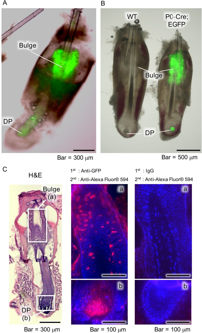
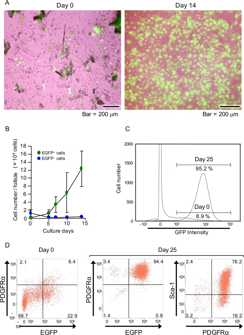
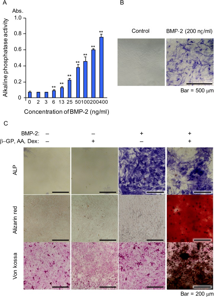
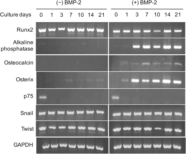
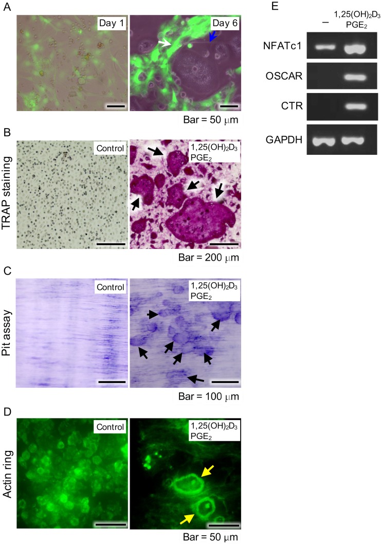
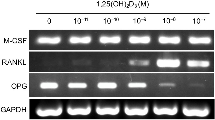
The neural crest (NC) arises near the neural tube during embryo development. NC cells migrate throughout the embryo and have potential to differentiate into multiple cell types, such as peripheral nerves, glial, cardiac smooth muscle, endocrine, and pigment cells, and craniofacial bone. In the present study, we induced osteoblast-like cells using whisker follicles obtained from the NC of mice. Hair follicle cells derived from the NC labeled with enhanced green fluorescent protein (EGFP) were collected from protein zero-Cre/floxed-EGFP double transgenic mice and cultured, then treated and cultured in stem cell growth medium. After growth for 14 days, results of flow cytometry analysis showed that 95% of the EGFP-positive (EGFP+) hair follicle cells derived from the NC had proliferated and 76.2% of those expressed mesenchymal stem cells markers, such as platelet-derived growth factor α and stem cell antigen-1, and also showed constitutive expression of Runx2 mRNA. Cells stimulated with bone morphogenetic protein-2 expressed osteocalcin, osterix, and alkaline phosphatase mRNA, resulting in production of mineralized matrices, which were detected by von Kossa and alizarin red staining. Moreover, EGFP+ hair follicle cells consistently expressed macrophage colony-stimulating factor and osteoprotegerin (OPG). Addition of 1α,25-dihydroxyvitamin D3 [1,25(OH)2D3] (10-8 M) to the cultures suppressed OPG expression and induced RANKL production in the cells. Furthermore, multinucleated osteoclasts appeared within 6 days after starting co-cultures of bone marrow cells with EGFP+ cells in the presence of 1,25(OH)2D3 and PGE2. These results suggest that NC-derived hair follicle cells possess a capacity for osteoblastic differentiation and may be useful for developing new bone regenerative medicine therapies.


Conflict of interest statement

Competing Interests: The authors have declared that no competing interests exist.

Figures

Fig 1
Fig 1. Distribution of NCDCs in whisker follicles.
(A) Fluorescence microscopy images of whisker follicle samples obtained from P0-Cre; CAG-CAT-EGFP Tg mice. Bulge, hair follicle bulge area; DP, dermal papilla. (B) Fluorescence microscopy images of whisker follicle samples obtained from wild-type (WT) and P0-Cre; CAG-CAT-EGFP Tg (P0-Cre; EGFP) mice. (C) Histological and immunohistochemical analysis findings of whisker follicles from P0-Cre; CAG-CAT-EGFP Tg mice following hematoxylin and eosin staining (left panel), and immunostaining with anti-GFP (middle panel), as well as of the negative control (right panel). Boxed areas in left panel indicate the bulge (a) and DP (b), which are shown at higher magnification in the middle and right panels.
Fig 2
Fig 2. Examination of proliferative potential and level of mesenchymal stem cells in NCDFCs.
(A) Phase-contrast images of whisker follicle cells subsequent to enzymatic processing on day 0 (left panel) and cells cultured in stem cell growth medium for 14 days (right panel). (B) The number of EGFP+ cells per whisker follicle increased with culture time. (C) EGFP-gated flow cytometry analysis charts of whisker follicle cells. EGFP+ cells were detected immediately after sampling (day 0) as well as after 25 days of proliferation in stem cell growth medium. (D) Flow cytometry analysis of cell-surface markers using freshly isolated whisker follicle cells from P0-Cre; CAG-CAT-EGFP Tg mice (day 0) and after 25 days of proliferation in stem cell growth medium. Representative flow cytometry images revealing EGFP- and PDGFRα-gated (left and middle panels) cells, and PDGFRα- and Sca-1-gated cells (right panel). Data shown represent mean values from 3 independent experiments, with error bars indicating SD.
Fig 3
Fig 3. Osteogenic differentiation of NCDFCs.
(A, B) Analysis of ALP expression. (A) ALP activity was elevated following addition of BMP-2 in a dose-dependent manner; Abs, absorbance. (B) Positive staining for ALP following addition of BMP-2. (C) Detection of calcification in calcification medium following alizarin red (middle panel), and von Kossa (lower panel) staining, and ALP expression (upper panel). β-GP, β-glycerophosphate; AA, ascorbic acid; Dex, dexamethasone. Data shown represent mean values from 3 independent experiments, with error bars indicating SD. **P<0.01.
Fig 4
Fig 4. Expression patterns of osteoblast-related and NC cell-related genes in NCDFCs.
Proliferative NCDFCs were stimulated with or without BMP-2, then mRNA expression levels were analyzed over time. All cells continuously expressed Runx2 mRNA, while stimulation with BMP-2 induced expressions of the osteoblastic differentiation-related genes ALP, osterix, and osteocalcin. In contrast, expression of p75, an NC cell-related gene, was not seen with or without BMP-2.
Fig 5
Fig 5. Examination of osteoblast functions supporting differentiation of osteoclasts.
(A) NCDFCs co-cultured with mouse bone-marrow cells including osteoclast precursor cells. Phase-contrast images of proliferative NCDFCs and mouse bone marrow cells in the presence of PGE2 and 1,25(OH)2D3 on day 1 (left panel). Formation of osteoclast-like cells (blue arrow in right panel) alongside EGFP+ cells (white arrow in right panel) on day 6. (B) Formation of osteoclasts in presence of PGE2 and 1,25(OH)2D3 detected by TRAP staining (arrows) after 6 days. (C) Cells showed bone resorption capacity after 6 days, as detected by toluidine blue staining (arrows). (D) Cells formed actin rings specific to the cytoskeleton of osteoclasts (yellow arrows), as detected by FITC-phalloidin staining after 6 days. (E) Semi quantitative RT-PCR measurements of expression of the osteoclast-related genes NFATc1, OSCAR, and calcitonin receptor in the presence of 1,25(OH)2D3 and PGE2. PGE2, prostaglandin E2; RT-PCR, reverse transcription-polymerase chain reaction.
Fig 6
Fig 6. Analysis of mRNA expression levels in proliferative NCDFCs stimulated with various concentrations of 1,25(OH)2D3.
RT-PCR analysis of NCDFCs exposed to osteoclast differentiation stimulating factors (M-CSF, RANKL) and an osteoclast differentiation inhibiting factor (OPG). Proliferative NCDFCs continuously expressed M-CSF. Addition of 1,25(OH)2D3 to the cultures suppressed the expression of OPG and induced production of RANKL by the cells.

Similar articles

Cited by

References

    1. Le Douarin NM. Cell migrations in embryos. Cell. 1984;38(2):353–60. - PubMed
    1. Lumsden A, Sprawson N, Graham A. Segmental origin and migration of neural crest cells in the hindbrain region of the chick embryo. Development. 1991;113(4):1281–91. - PubMed
    1. Chung KF, Sicard F, Vukicevic V, Hermann A, Storch A, Huttner WB, et al. Isolation of neural crest derived chromaffin progenitors from adult adrenal medulla. Stem Cells. 2009;27(10):2602–13. doi: 10.1002/stem.180 - DOI - PubMed
    1. Osumi-Yamashita N, Ninomiya Y, Doi H, Eto K. The contribution of both forebrain and midbrain crest cells to the mesenchyme in the frontonasal mass of mouse embryos. Dev Biol. 1994;164(2):409–19. doi: 10.1006/dbio.1994.1211 - DOI - PubMed
    1. Chai Y, Jiang X, Ito Y, Bringas P Jr., Han J, Rowitch DH, et al. Fate of the mammalian cranial neural crest during tooth and mandibular morphogenesis. Development. 2000;127(8):1671–9. Epub 2000/03/22. - PubMed