. 2017 Apr 6;12(4):e0173924.
doi: 10.1371/journal.pone.0173924. eCollection 2017.

Transmembrane domain quality control systems operate at the endoplasmic reticulum and Golgi apparatus

Affiliations

Transmembrane domain quality control systems operate at the endoplasmic reticulum and Golgi apparatus

Kit Briant et al. PLoS One. .

Abstract

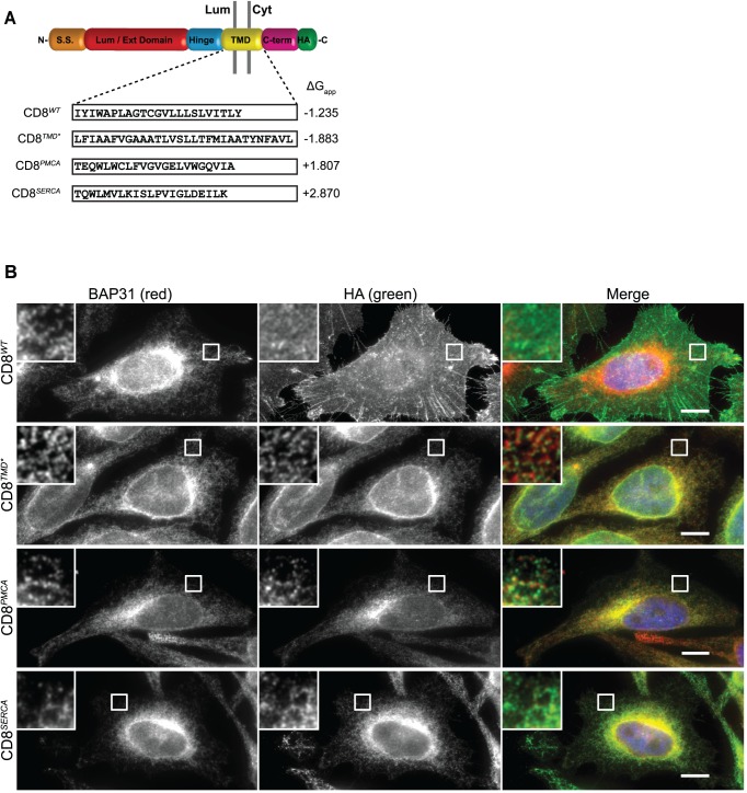
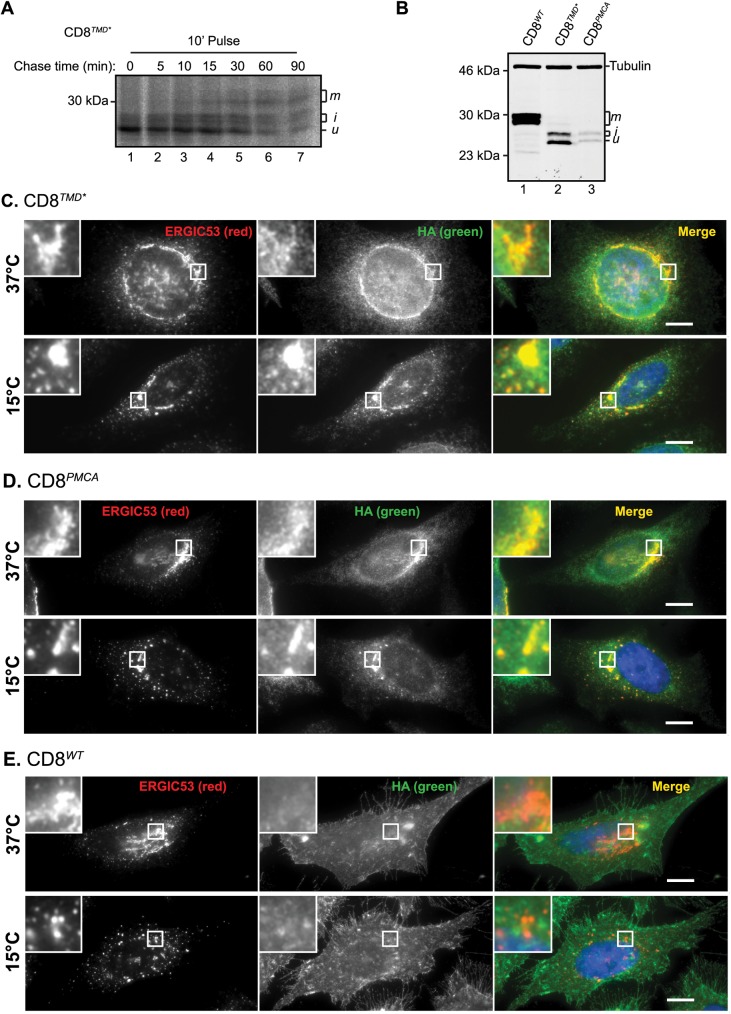
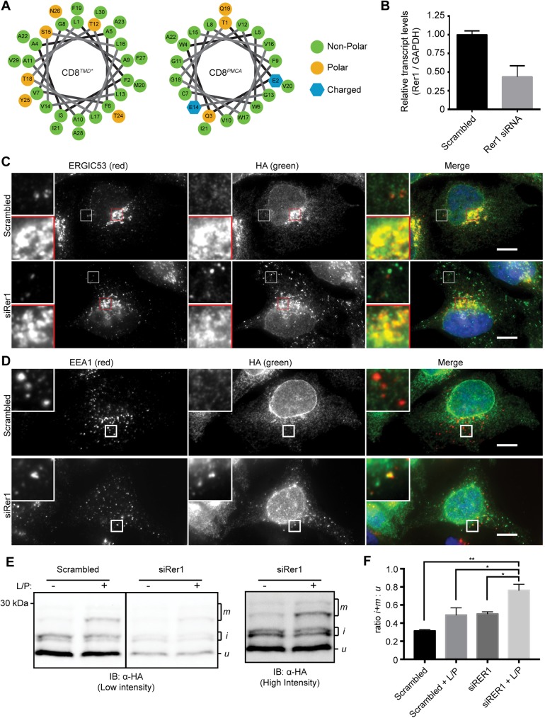
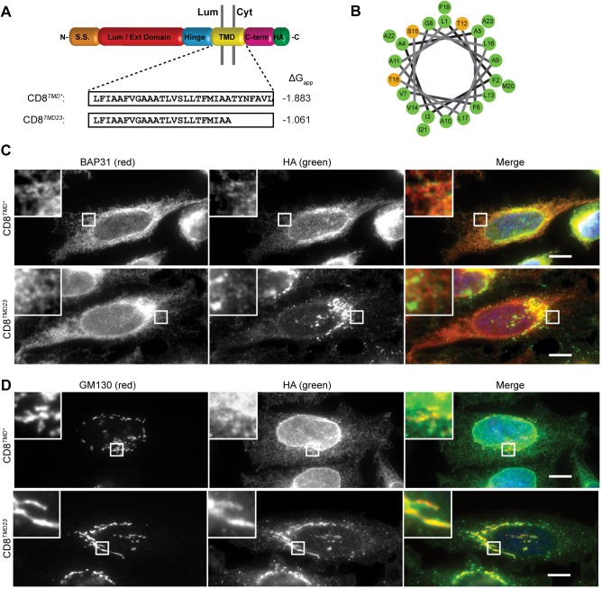
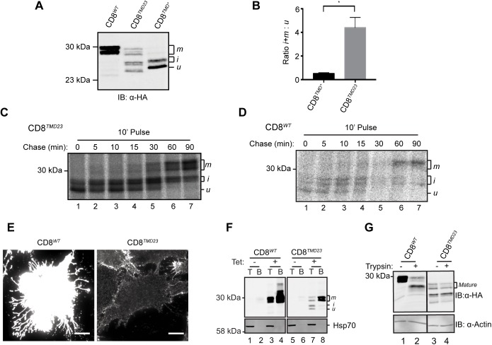
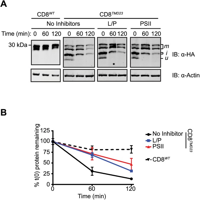
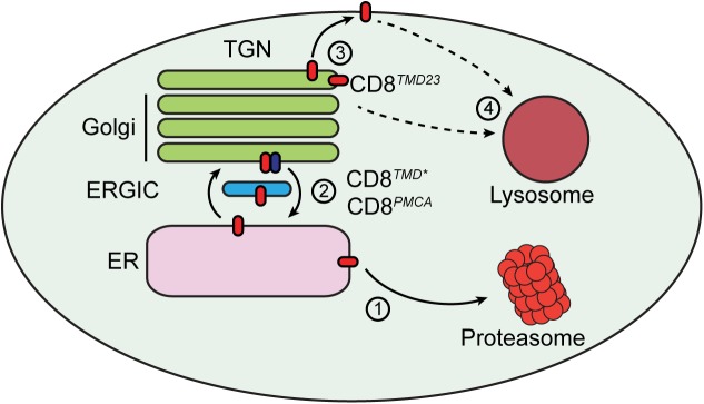
Multiple protein quality control systems operate to ensure that misfolded proteins are efficiently cleared from the cell. While quality control systems that assess the folding status of soluble domains have been extensively studied, transmembrane domain (TMD) quality control mechanisms are poorly understood. Here, we have used chimeras based on the type I plasma membrane protein CD8 in which the endogenous TMD was substituted with transmembrane sequences derived from different polytopic membrane proteins as a mode to investigate the quality control of unassembled TMDs along the secretory pathway. We find that the three TMDs examined prevent trafficking of CD8 to the cell surface via potentially distinct mechanisms. CD8 containing two distinct non-native transmembrane sequences escape the ER and are subsequently retrieved from the Golgi, possibly via Rer1, leading to ER localisation at steady state. A third chimera, containing an altered transmembrane domain, was predominantly localised to the Golgi at steady state, indicating the existence of an additional quality control checkpoint that identifies non-native transmembrane domains that have escaped ER retention and retrieval. Preliminary experiments indicate that protein retained by quality control mechanisms at the Golgi are targeted to lysosomes for degradation.


Conflict of interest statement

Competing Interests: The authors have declared that no competing interests exist.

Figures

Fig 1
Fig 1. Diverse unassembled TMDs mediate ER localisation.
(A) Cartoon schematic of the domain structure of CD8α. The TMD amino acid sequence and predicted ΔGapp for TMD membrane insertion of CD8WT, CD8TMD*, CD8PMCA and CD8SERCA are shown. (B) Cells expressing CD8WT, CD8TMD*, CD8PMCA or CD8SERCA were fixed with formaldehyde, permeabilised with Triton X-100 and co-immunostained with antibodies against BAP31 and the epitope tag HA. Cell nuclei were stained with DAPI (represented in blue in merged images). Scale bars indicate 10 μm.
Fig 2
Fig 2. CD8TMD* and CD8PMCA are retrieved from the Golgi.
(A) Cells expressing CD8TMD* were pulse labelled with [35S] met/cys for 10 minutes and chased for up to 90 minutes as indicated. Samples were immunoprecipitated with antibodies against HA, separated by SDS-PAGE and analysed by phosphorimaging. (B) Whole cell lysates of cells expressing CD8WT, CD8TMD* or CD8PMCA were separated by SDS-PAGE and analysed by immunoblotting with antibodies against HA and tubulin. (C-E) Cells expressing (C) CD8TMD*, (D) CD8PMCA or (E) CD8WT were incubated at 15°C or 37°C for 3 hours prior fixation. Cells were co-immunostained with antibodies against ERGIC53 and the epitope tag HA. Scale bars indicate 10 μm.
Fig 3
Fig 3. CD8TMD* retrieval from the Golgi is mediated by Rer1.
(A) Helical wheel projections for the transmembrane domains of CD8TMD* and CD8PMCA. Numbering starts from the N-terminus of the predicted TMD. (B) Rer1 mRNA levels were determined by qPCR 72h post transfection with scrambled or Rer1 siRNA and normalised relative to GAPDH mRNA levels. (C and D) Cells expressing CD8TMD* were transfected with scrambled siRNA or siRNA targeting Rer1 as indicated. 72 hours subsequently, cells were fixed and co-immunostained with antibodies against (C) ERGIC53 and HA or (D) EEA1 and HA. Cell nuclei were stained with DAPI. Scale bars indicate 10 μm. (E) Cell expressing CD8TMD* were transfected with scrambled siRNA or siRNA targeting Rer1 for 72 hours. Immediately prior to harvesting, cells were incubated with leupeptin and pepstatin A (L/P) for 5 hours or left uninhibited. Whole cell lysates were separated by SDS-PAGE and analysed by immunoblotting with antibodies against HA. (F) Signal intensities from high molecular weight ‘i’ and ‘m’ glycoforms expressed as a ratio to the unprocessed ‘u’ glycoform. Scrambled siRNA vs RER1 siRNA + L/P, p = 0.0015; Rer1 siRNA vs Rer1 siRNA + L/P, p = 0.0267; scrambled siRNA + L/P vs Rer1 siRNA + L/P, p = 0.0337; one way ANOVA with Tukey’s multiple comparisons test. Data represents mean ±S.E.M. from 3 independent experiments.
Fig 4
Fig 4. CD8TMD23 is a substrate from Golgi quality control.
(A) Cartoon schematic of the domain structure of CD8α. The amino acid compositions and predicted ΔGapp for TMD membrane insertion of CD8TMD* and CD8TMD23 are shown. (B) Helical wheel projection for the transmembrane domain of CD8TMD23. Numbering starts from the N-terminus of the predicted TMD. (C and D) Cells expressing CD8TMD* or CD8TMD23 were fixed and co-immunostained with antibodies against (C) BAP31 or (D) GM130 as well as the epitope tag HA in all cases. Cell nuclei were stained with DAPI. Scale bars indicate 10 μm.
Fig 5
Fig 5. CD8TMD23 is predominantly retained in the Golgi.
(A) Whole cell lysates of cells expressing CD8WT, CD8TMD* or CD8TMD23 were separated by SDS-PAGE and analysed by immunoblotting with antibodies against HA. (B) Ratios of signal intensities of intermediate ‘i’ and mature ‘m’ to unprocessed ‘u’ glycoforms of CD8TMD* or CD8TMD23 at steady state (n = 3). P = 0.0109, unpaired t-test. (C & D) Cells expressing (C) CD8TMD23 or (D) CD8WT were pulse labelled with [35S] met/cys for 10 minutes and chased for up to 90 minutes as indicated. Samples were immunoprecipitated with antibodies against HA, separated by SDS-PAGE and analysed by phosphorimaging. (E) In parallel, cells expressing CD8WT or CD8TMD23 were chilled on ice and labelled with antibodies against the extracellular domain of CD8 prior to fixation in formaldehyde. Note that pictures were taken in parallel with equal exposure times. Scale bars indicate 10 μm. (F) Cells expressing CD8WT or CD8TMD23 were chilled on iced water and labelled with biotin prior to cell lysis. Total cell lysates samples were taken and the remaining cell lysate incubated with neutravidin to isolate biotinylated (cell surface) protein. Total ‘T’ and biotinylated ‘B’ samples were separated by SDS-PAGE and analysed by immunoblotting with antibodies against HA, tubulin and Hsp70. (G) Cells expressing CD8WT or CD8TMD23 were incubated on ice and treated with trypsin. Subsequently cells were washed and whole cell lysates separated by SDS-PAGE and analysed by immunoblotting with antibodies against HA and actin.
Fig 6
Fig 6. CD8TMD23 is degraded by proteasomes and lysosomes.
(A) Cells expressing CD8WT or CD8TMD23 were treated with cycloheximide (CHX) to prevent further protein synthesis. Whole cell lysates were harvested at the indicated chase time, separated by SDS-PAGE and analysed by immunoblotting with antibodies against HA and the loading control actin. Where indicated, cells expressing CD8TMD23 were treated with leupeptin and pepstatin A (LP) or PSII concurrently with CHX. (B) Signal intensities from (A) were quantified, normalised relative to the loading control and expressed as a percentage of the protein level at the start of the chase. Data represents mean ±S.E.M. from 3 independent experiments.
Fig 7
Fig 7. Transmembrane domain quality control at the ER and Golgi.
A model for the handling of proteins containing misassembled transmembrane domains (red ovals). (1) Proteins containing misassembled transmembrane domains may be retained in the ER and degraded via ERAD. (2) A proportion of these proteins may cycle between the ER and Golgi. Golgi-to-ER retrieval is at least in part mediated by Rer1 (blue oval). (3) Proteins that are not retrieved to the ER may be retained by Golgi quality control machinery. (4) Proteins that escape ER retrieval are degraded in lysosomes. Some protein may traffic to the cell surface. It is not clear whether trafficking to the plasma membrane is a prerequisite for targeting to lysosomes, or whether proteins containing aberrant TMDs can be directly targeted to the endo/lysosomal system from the Golgi.

Similar articles

Cited by

References

    1. Wallin E, von Heijne G. Genome-wide analysis of integral membrane proteins from eubacterial, archaean, and eukaryotic organisms. Protein Sci. 1998;7(4):1029–38. Epub 1998/05/06. PubMed Central PMCID: PMCPmc2143985. doi: 10.1002/pro.5560070420 - DOI - PMC - PubMed
    1. Braakman I, Hebert DN. Protein folding in the endoplasmic reticulum. Cold Spring Harb Perspect Biol. 2013;5(5):a013201 Epub 2013/05/03. doi: 10.1101/cshperspect.a013201 - DOI - PMC - PubMed
    1. Guo Y, Sirkis DW, Schekman R. Protein Sorting at the trans-Golgi network. Annu Rev Cell Dev Biol. 2014;30:169–206. Epub 2014/08/26. doi: 10.1146/annurev-cellbio-100913-013012 - DOI - PubMed
    1. Babst M. Quality control: quality control at the plasma membrane: one mechanism does not fit all. J Cell Biol. 2014;205(1):11–20. Epub 2014/04/16. PubMed Central PMCID: PMCPmc3987138. doi: 10.1083/jcb.201310113 - DOI - PMC - PubMed
    1. Koenig PA, Ploegh HL. Protein quality control in the endoplasmic reticulum. F1000Prime Rep. 2014;6:49 Epub 2014/09/04. PubMed Central PMCID: PMCPmc4108957. doi: 10.12703/P6-49 - DOI - PMC - PubMed