. 2017 Apr 5;94(1):48-57.e4.
doi: 10.1016/j.neuron.2017.03.027.

Polyglutamine-Expanded Huntingtin Exacerbates Age-Related Disruption of Nuclear Integrity and Nucleocytoplasmic Transport

Affiliations

Polyglutamine-Expanded Huntingtin Exacerbates Age-Related Disruption of Nuclear Integrity and Nucleocytoplasmic Transport

Fatima Gasset-Rosa et al. Neuron. .

Abstract

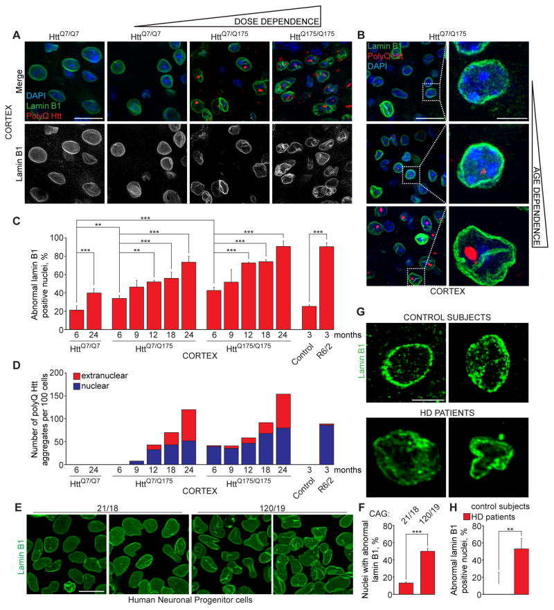
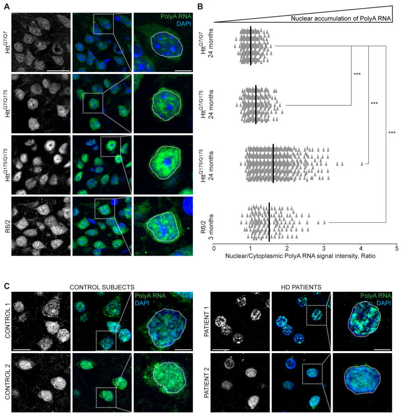
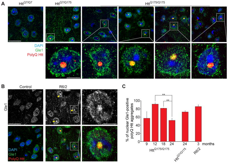
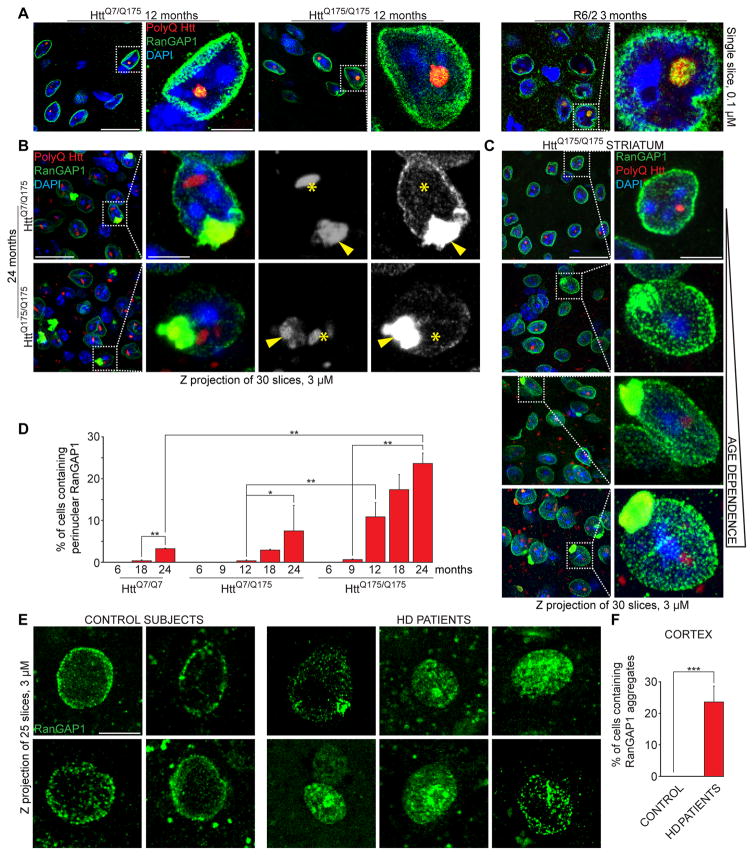
Onset of neurodegenerative disorders, including Huntington's disease, is strongly influenced by aging. Hallmarks of aged cells include compromised nuclear envelope integrity, impaired nucleocytoplasmic transport, and accumulation of DNA double-strand breaks. We show that mutant huntingtin markedly accelerates all of these cellular phenotypes in a dose- and age-dependent manner in cortex and striatum of mice. Huntingtin-linked polyglutamine initially accumulates in nuclei, leading to disruption of nuclear envelope architecture, partial sequestration of factors essential for nucleocytoplasmic transport (Gle1 and RanGAP1), and intranuclear accumulation of mRNA. In aged mice, accumulation of RanGAP1 together with polyglutamine is shifted to perinuclear and cytoplasmic areas. Consistent with findings in mice, marked alterations in nuclear envelope morphology, abnormal localization of RanGAP1, and nuclear accumulation of mRNA were found in cortex of Huntington's disease patients. Overall, our findings identify polyglutamine-dependent inhibition of nucleocytoplasmic transport and alteration of nuclear integrity as a central component of Huntington's disease.


Figures

Figure 1
Figure 1. Expanded polyQ Huntingtin disrupts neuronal nuclear envelope morphology in a dose- and age-dependent manner
(A) Immunofluorescence (IF) of lamin B1 (green) and polyQ Htt (red) in cortex of mice at indicated ages expressing one (HttQ7/Q175) or two (HttQ175/Q175) copies of expanded polyQ Htt and wild type littermates (HttQ7/Q7). (B) IF of lamin B1 (green) and polyQ Htt (red) in cortex of aging HttQ7/Q175 mice. A, B: Nuclei were stained with DAPI. (C) Percentage of altered nuclear envelopes in cortical sections of mice of indicated genotypes and ages, as determined by IF of lamin B1. 100–400 nuclei were counted per genotype and age. (D) Quantification of nuclear and extranuclear aggregates of polyQ Htt in cortex of HttQ175 and R6/2 mice. 200 cells were counted per genotype and age. (E, F) IF of lamin B1 (E) and percentage of altered nuclear envelopes (F) in human iPSC-derived neuronal progenitor cells with indicated number of CAG repeats. At least 600 cells were analyzed for each genotype. (G, H) IF of lamin B1 (G) and percentage of altered nuclear envelopes (H) in sections of motor cortex of two non-neurological disease control subjects and two Huntington’s disease patients of indicated ages. At least 200 nuclei were counted per each group. A, B, G: Z projections (3 μM) are shown. C, F, H: Data are shown as mean ± SEM. **: P<0.01, ***: P<0.001, chi-square test. See also Figures S1 and S2.
Figure 2
Figure 2. Expanded polyQ Huntingtin impairs mRNA export from nuclei
(A) Fluorescence in situ hybridization (FISH) of polyA RNA (green) of cortex from mice of indicated genotypes and ages. Z projections of 30 consecutive slices (3 μM) are shown. (B) Quantification of (A). The ratio of nuclear to cytoplasmic signal intensities of PolyA RNA per cell in cortex from mice of indicated genotypes and ages normalized by 24 months HttQ7/Q7 mean. Each individual point represents a single cell. Mean is shown per each genotype and age. ***: P<0.001, unpaired t test, comparison with control 24 months HttQ7/Q7. 300–600 cells were counted per genotype and age. (C) FISH of polyA RNA (green) in sections of motor cortex of non-neurological disease control subjects and Huntington’s disease patients. Z projections of 30 consecutive slices (3 μM) are shown. A, C: nuclei were stained with DAPI. See also Figure S3.
Figure 3
Figure 3. Nuclear polyQ Htt aggregates co-localize with the mRNA export factor Gle1
(A, B) Immunofluorescence of polyQ Htt (red) and Gle1 (green) in cortical sections of (A) HttQ7/Q7, HttQ7/Q175 and HttQ175/Q175 mice of indicated ages and (B) 3-month-old control and R6/2 mice. Nuclei were stained with DAPI. (C) Percentage of nuclear polyQ aggregates positive for Gle1, in mice of indicated genotypes and ages. Data are shown as mean ± SEM. **: P<0.01, chi-square test. 100 cells were counted per genotype and age. A, B: Arrowheads point to Gle1/polyQ Htt nuclear co-localization. See also Figure S4.
Figure 4
Figure 4. Expanded polyQ Huntingtin sequesters RanGAP1
(A) Immunofluorescence (IF) of RanGAP1 (green) and polyQ Htt (red) in cortical sections of mice of indicated genotypes and ages. Arrowheads point to RanGap1/polyQ Htt nuclear co-localization. Single slices (0.1 μM) are shown. (B) IF of RanGAP1 (green) and polyQ Htt (red) in cortex of 24-month old mice of indicated genotypes. Localization of nuclear (asterisks) and perinuclear (arrowheads) polyQ Htt aggregates is shown. (C) IF of RanGAP1 in striatal sections of HttQ175/Q175 mice of indicated ages. (D) Percentage of cells containing perinuclear RanGAP1 in cortical sections of mice of indicated genotypes and ages. At least 600 cells per animal were analyzed. (E, F) IF of RanGAP1 (E) and percentage of cells containing mislocalized RanGAP1 (F) in sections of motor cortex of non-neurological disease control subjects and Huntington’s disease patients. At least 200 nuclei were counted per each group. B, C, E: Z projections (3 μM) are shown. D, F: Data are shown as mean ± SEM. *: P<0.05, **: P<0.01, ***: P<0.001, chi-square test. See also Figure S4.

References

    1. Bennett EJ, Shaler TA, Woodman B, Ryu KY, Zaitseva TS, Becker CH, Bates GP, Schulman H, Kopito RR. Global changes to the ubiquitin system in Huntington’s disease. Nature. 2007;448:704–708. - PubMed
    1. Bischoff FR, Krebber H, Kempf T, Hermes I, Ponstingl H. Human RanGTPase-activating protein RanGAP1 is a homologue of yeast Rna1p involved in mRNA processing and transport. Proc Natl Acad Sci U S A. 1995;92:1749–1753. - PMC - PubMed
    1. Bukata L, Parker SL, D’angelo MA. Nuclear pore complexes in the maintenance of genome integrity. Curr Opin Cell Biol. 2013;25:378–386. - PubMed
    1. Carty N, Berson N, Tillack K, Thiede C, Scholz D, Kottig K, Sedaghat Y, Gabrysiak C, Yohrling G, Von Der Kammer H, et al. Characterization of HTT inclusion size, location, and timing in the zQ175 mouse model of Huntington’s disease: an in vivo high-content imaging study. PLoS One. 2015;10:e0123527. - PMC - PubMed
    1. Chapple JP, Bros-Facer V, Butler R, Gallo JM. Focal distortion of the nuclear envelope by huntingtin aggregates revealed by lamin immunostaining. Neurosci Lett. 2008;447:172–174. - PMC - PubMed

Publication types

MeSH terms