

Regulation of the JMJD3 (KDM6B) histone demethylase in glioblastoma stem cells by STAT3
- PMID: 28384648
- PMCID: PMC5383422
- DOI: 10.1371/journal.pone.0174775
Regulation of the JMJD3 (KDM6B) histone demethylase in glioblastoma stem cells by STAT3
Abstract
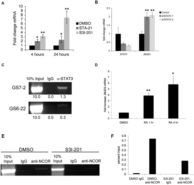
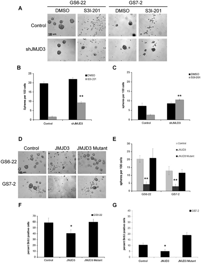
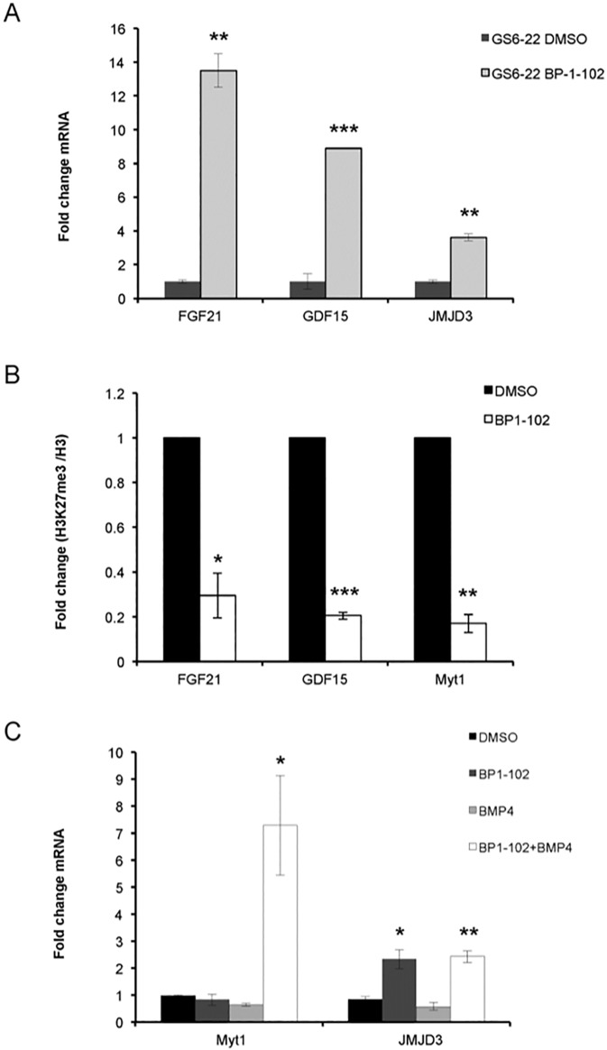
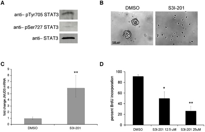
The growth factor and cytokine regulated transcription factor STAT3 is required for the self-renewal of several stem cell types including tumor stem cells from glioblastoma. Here we show that STAT3 inhibition leads to the upregulation of the histone H3K27me2/3 demethylase Jmjd3 (KDM6B), which can reverse polycomb complex-mediated repression of tissue specific genes. STAT3 binds to the Jmjd3 promoter, suggesting that Jmjd3 is a direct target of STAT3. Overexpression of Jmjd3 slows glioblastoma stem cell growth and neurosphere formation, whereas knockdown of Jmjd3 rescues the STAT3 inhibitor-induced neurosphere formation defect. Consistent with this observation, STAT3 inhibition leads to histone H3K27 demethylation of neural differentiation genes, such as Myt1, FGF21, and GDF15. These results demonstrate that the regulation of Jmjd3 by STAT3 maintains repression of differentiation specific genes and is therefore important for the maintenance of self-renewal of normal neural and glioblastoma stem cells.
Conflict of interest statement
Figures
Similar articles
-
The role and prospect of JMJD3 in stem cells and cancer.Biomed Pharmacother. 2019 Oct;118:109384. doi: 10.1016/j.biopha.2019.109384. Epub 2019 Sep 6. Biomed Pharmacother. 2019. PMID: 31545292 Review.
-
The signal transducers Stat1 and Stat3 and their novel target Jmjd3 drive the expression of inflammatory genes in microglia.J Mol Med (Berl). 2014 Mar;92(3):239-54. doi: 10.1007/s00109-013-1090-5. Epub 2013 Oct 6. J Mol Med (Berl). 2014. PMID: 24097101 Free PMC article.
-
JMJD3 suppresses stem cell-like characteristics in breast cancer cells by downregulation of Oct4 independently of its demethylase activity.Oncotarget. 2017 Mar 28;8(13):21918-21929. doi: 10.18632/oncotarget.15747. Oncotarget. 2017. PMID: 28423536 Free PMC article.
-
The histone demethylase jumonji coordinates cellular senescence including secretion of neural stem cell-attracting cytokines.Mol Cancer Res. 2015 Apr;13(4):636-50. doi: 10.1158/1541-7786.MCR-13-0268. Epub 2015 Feb 4. Mol Cancer Res. 2015. PMID: 25652587 Free PMC article.
-
JMJD3 as an epigenetic regulator in development and disease.Int J Biochem Cell Biol. 2015 Oct;67:148-57. doi: 10.1016/j.biocel.2015.07.006. Epub 2015 Jul 17. Int J Biochem Cell Biol. 2015. PMID: 26193001 Free PMC article. Review.
Cited by
-
JMJD family proteins in cancer and inflammation.Signal Transduct Target Ther. 2022 Sep 1;7(1):304. doi: 10.1038/s41392-022-01145-1. Signal Transduct Target Ther. 2022. PMID: 36050314 Free PMC article. Review.
-
Emerging roles of JMJD3 in cancer.Clin Transl Oncol. 2022 Jul;24(7):1238-1249. doi: 10.1007/s12094-021-02773-9. Epub 2022 Mar 3. Clin Transl Oncol. 2022. PMID: 35239138 Review.
-
Epigenetic regulation of macrophage polarization in wound healing.Burns Trauma. 2023 Jan 17;11:tkac057. doi: 10.1093/burnst/tkac057. eCollection 2023. Burns Trauma. 2023. PMID: 36687556 Free PMC article. Review.
-
The JMJD Family Histone Demethylases in Crosstalk Between Inflammation and Cancer.Front Immunol. 2022 Apr 26;13:881396. doi: 10.3389/fimmu.2022.881396. eCollection 2022. Front Immunol. 2022. PMID: 35558079 Free PMC article. Review.
-
KDM6B promotes activation of the oncogenic CDK4/6-pRB-E2F pathway by maintaining enhancer activity in MYCN-amplified neuroblastoma.Nat Commun. 2021 Dec 10;12(1):7204. doi: 10.1038/s41467-021-27502-2. Nat Commun. 2021. PMID: 34893606 Free PMC article.
References
-
- Lee J, Kotliarova S, Kotliarov Y, Li A, Su Q, Donin NM, et al. Tumor stem cells derived from glioblastomas cultured in bFGF and EGF more closely mirror the phenotype and genotype of primary tumors than do serum-cultured cell lines. Cancer Cell. 2006;9(5):391–403. doi: 10.1016/j.ccr.2006.03.030 - DOI - PubMed
-
- Vescovi AL, Galli R, Reynolds BA. Brain tumour stem cells. Nature reviews. 2006;6(6):425–36. doi: 10.1038/nrc1889 - DOI - PubMed
-
- Yu H, Jove R. The STATs of cancer—new molecular targets come of age. Nature reviews. 2004;4(2):97–105. doi: 10.1038/nrc1275 - DOI - PubMed
-
- Darnell JE. Validating Stat3 in cancer therapy. Nat Med. 2005;11(6):595–6. doi: 10.1038/nm0605-595 - DOI - PubMed
MeSH terms
Substances
Grants and funding
LinkOut - more resources
Full Text Sources
Other Literature Sources
Medical
Molecular Biology Databases
Research Materials
Miscellaneous
