. 2017 Jun;23(7):1035-1039.
doi: 10.1177/1352458516679035. Epub 2017 Apr 7.

Retrograde trans-synaptic visual pathway degeneration in multiple sclerosis: A case series

Affiliations

Retrograde trans-synaptic visual pathway degeneration in multiple sclerosis: A case series

Omar Al-Louzi et al. Mult Scler. 2017 Jun.

Abstract

Background: Trans-synaptic degeneration (TSD) describes the propagation of neuronal injury through synaptic pathways in the human nervous system and may be linked to the accelerated retinal atrophy seen in multiple sclerosis (MS).

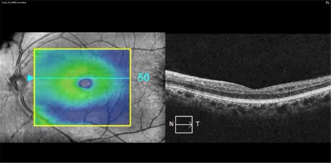
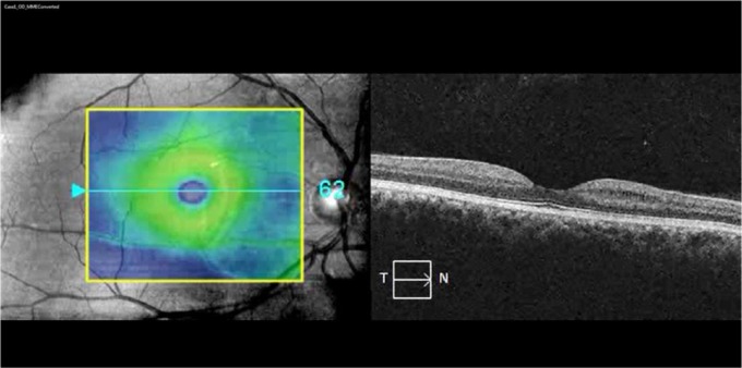
Results: We report six cases where homonymous, hemi-macular ganglion cell + inner plexiform (GCIP) thickness reduction was seen in conjunction with posterior visual pathway lesions. Macular microcystoid changes of the inner nuclear layer (INL) were seen in a subset of three subjects.

Conclusion: Our findings highlight the utility of assessing regional GCIP changes to identify potential retrograde TSD in MS and demonstrate that INL changes may be an accompaniment in such instances.

Keywords: MRI; Relapsing/remitting; T2 lesions; atrophy; optical coherence tomography; retina.


Conflict of interest statement

Declaration of Conflicting Interests: The author(s) declared the following potential conflicts of interest with respect to the research, authorship, and/or publication of this article: Dr Al-Louzi and J. Button report no disclosures. Dr Newsome has received consultant fees for scientific advisory boards from Biogen, Genzyme, and Novartis and has received research funding from Biogen and Novartis. Dr Calabresi has received personal compensation for consulting and serving on scientific advisory boards from Vertex, Vaccinex, Merck, and AbbVie and has received research funding from Biogen-Idec, MedImmune, and Novartis and has also received National Institutes of Health grant 5R01NS082347-02. Dr Saidha has received consulting fees from Medical Logix for the development of CME programs in neurology, consulting fees from Axon Advisors LLC, Educational Grant Support from Novartis and Teva Neurosciences, speaking honoraria from the National Association of Managed Care Physicians, Family Medicine Foundation of West Virginia, and Advanced Studies in Medicine and served on scientific advisory boards for Biogen-Idec, Genzyme, and Novartis. He also received research funding from the Race to Erase MS and receives an unrestricted research grant from Genentech Corporation.

Figures

Figure 1.
Figure 1.
MRI and OCT findings: (a) case 1: FLAIR imaging showing a lesion in the left occipital white matter (red inset, arrow) associated with focal volume loss (red inset, arrowheads). OCT imaging revealed homonymous, hemi-macular GCIP thinning and MMP (red insets); (b) case 2: MPRAGE image showing a T1-hypointense lesion in the right occipital white matter extending into the adjacent striate cortex, which appears atrophic relative to the contralateral side (red inset). The lesion appeared T2 hyperintense on FLAIR imaging (green inset), while OCT showed homonymous, hemi-macular GCIP thinning and MMP; and (c) case 4: FLAIR image showing a right periventricular T2 hyperintensity extending into the occipital white matter in a patient who presented with left homonymous hemianopia. OCT images performed 4 years after presentation revealed hemi-macular GCIP thickness reduction without evidence of MMP. FLAIR: fluid-attenuated inversion recovery; OCT: optical coherence tomography; GCIP: ganglion cell + inner plexiform layers; MMP: macular microcystoid pathology; MPRAGE: magnetization-prepared rapid-acquisition with gradient echo; N: nasal; T: temporal.
None
None

Comment in

References

    1. Jindahra P, Petrie A, Plant GT. The time course of retrograde trans-synaptic degeneration following occipital lobe damage in humans. Brain 2012; 135(Pt 2): 534–541. - PubMed
    1. Klistorner A, Sriram P, Vootakuru N, et al. Axonal loss of retinal neurons in multiple sclerosis associated with optic radiation lesions. Neurology 2014; 82: 2165–2172. - PMC - PubMed
    1. Gabilondo I, Martínez-Lapiscina EH, Martínez-Heras E, et al. Trans-synaptic axonal degeneration in the visual pathway in multiple sclerosis. Ann Neurol 2014; 75(1): 98–107. - PubMed
    1. Oh J, Sotirchos ES, Saidha S, et al. In vivo demonstration of homonymous hemimacular loss of retinal ganglion cells due to a thalamic lesion using optical coherence tomography. JAMA Neurol 2013; 70(3): 410–411. - PMC - PubMed
    1. Tur C, Goodkin O, Altmann DR, et al. Longitudinal evidence for anterograde trans-synaptic degeneration after optic neuritis. Brain 2016; 139(3): 816–828. - PMC - PubMed

Publication types

MeSH terms

LinkOut - more resources