

Successful treatment with bortezomib and dexamethasone for proliferative glomerulonephritis with monoclonal IgG deposits in multiple myeloma: a case report
- PMID: 28385149
- PMCID: PMC5382661
- DOI: 10.1186/s12882-017-0524-7
Successful treatment with bortezomib and dexamethasone for proliferative glomerulonephritis with monoclonal IgG deposits in multiple myeloma: a case report
Abstract
Background: Proliferative glomerulonephritis with monoclonal IgG deposits (PGNMID) is a form of renal involvement by monoclonal IgG deposits that was found in mesangial, subendothelial or subepithelial regions. The distribution of glomerular deposits was completely different from that in monoclonal immunoglobulin deposition disease. PGNMID is reported to be rarely associated with a hematological malignancy. Previously, only five cases of PGNMID with multiple myeloma have been reported. However, the pathogenic relationship between PGNMID and multiple myeloma was unclear because a detailed description was not provided. We report that a patient with PGNMID associated with multiple myeloma was treated with bortezomib and dexamethasone and underwent the second renal biopsy after treatment, showing that chemotherapy was effective for PGNMID clinically and pathologically.
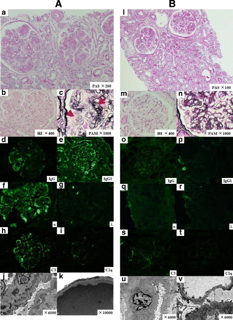
Case presentation: A 75-year-old man presented with progressive leg edema, had nephrotic range proteinuria, hypoalbuminemia, moderate renal failure, and occult blood in his urine. Electrophoresis results showed serum and urinary monoclonal spikes of IgGκ type immunoglobulin. A renal biopsy specimen showed lobular mesangial proliferation with mesangiolysis, glomerular micro-aneurysm, and endocapillary hypercellularity. Immunofluorescence results revealed strong granular capillary and mesangial staining for IgG1, C3 and κ light chain in glomeruli without tubular deposits of any immunoglobulin. Electron microscopy also showed dense granular deposits in subendothelial and mesangial areas. PGNMID associated with multiple myeloma (IgGκ type) was diagnosed on the basis of a subsequent bone marrow examination. Bortezomib and dexamethasone therapy significantly reduced proteinuria and elevated serum albumin level. Eight months later, the second renal biopsy showed no active lesions and that the IgG1 and κ light chain deposits had drastically disappeared.
Conclusions: This is the first case of PGNMID with multiple myeloma successfully treated with bortezomib and dexamethasone in which comparative renal biopsies were performed before and after treatment. Our findings suggest the pathogenesis of PGNMID and therapeutic options for PGNMID.
Keywords: Monoclonal gammopathy; Multiple myeloma; Proliferative glomerulonephritis with monoclonal IgG deposits (PGNMID).
Figures

References
-
- Nasr SH, Markowitz GS, Stokes MB, Seshan SV, Valderrama E, Appel GB, Aucouturier P, D’Agati VD. Proliferative glomerulonephritis with monoclonal IgG deposits: A distinct entity mimicking immune-complex glomerulonephritis. Kidney Int. 2004;65(1):85–96. doi: 10.1111/j.1523-1755.2004.00365.x. - DOI - PubMed
-
- Ranghino A, Tamagnone M, Messina M, Barreca A, Biancone L, Basolo B, Segoloni GP, Mazzucco G. A case of recurrent proliferative glomerulonephritis with monoclonal IgG deposits after kidney transplant treated with plasmapheresis. Case Rep Nephrol Urol. 2012;2(1):46–52. doi: 10.1159/000339405. - DOI - PMC - PubMed
Publication types
MeSH terms
Substances
LinkOut - more resources
Full Text Sources
Other Literature Sources
Medical
Miscellaneous
