. 2017 Jun 1;312(6):L1018-L1028.
doi: 10.1152/ajplung.00067.2017. Epub 2017 Apr 6.

Free actin impairs macrophage bacterial defenses via scavenger receptor MARCO interaction with reversal by plasma gelsolin

Affiliations

Free actin impairs macrophage bacterial defenses via scavenger receptor MARCO interaction with reversal by plasma gelsolin

Christine M Ordija et al. Am J Physiol Lung Cell Mol Physiol. .

Abstract

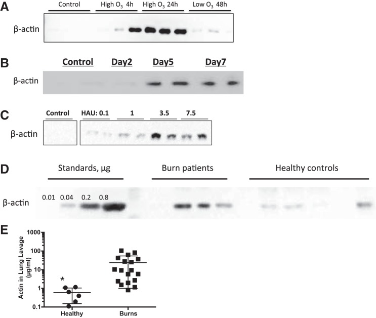
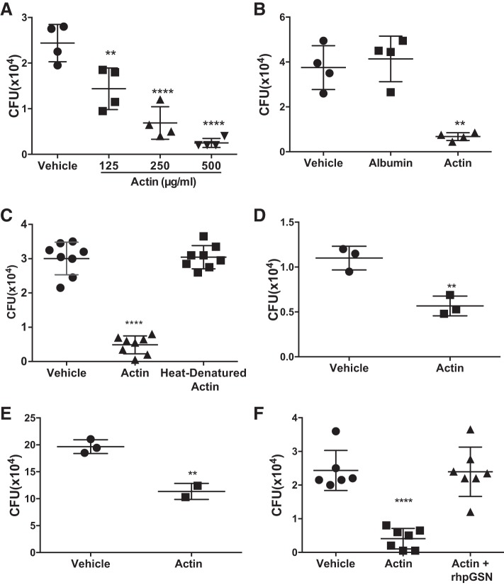
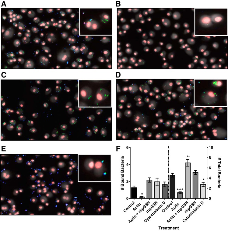
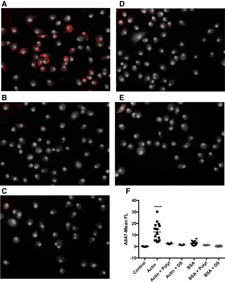
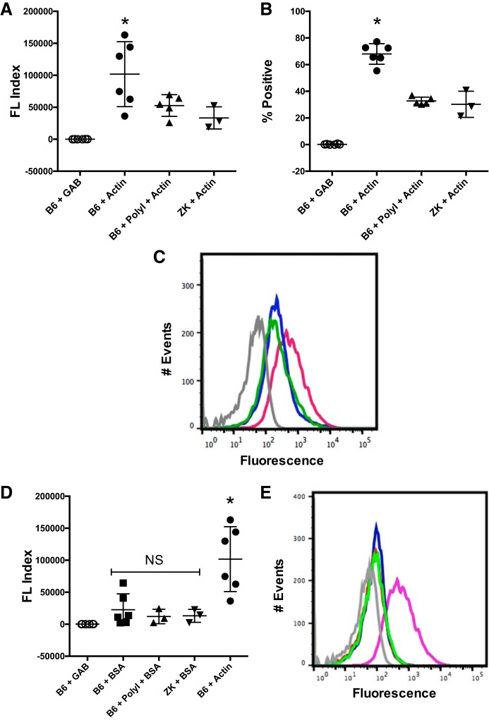
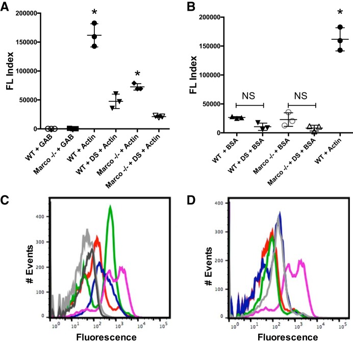
Lung injury can release intracellular actin into the alveolar milieu and is also associated with increased susceptibility to secondary infections. We investigated the effect of free (extracellular) actin on lung macrophage host defense functions. Western blot analysis demonstrated free actin release into the lung lavage fluids of mouse models of ozone injury, influenza infection, and secondary pneumococcal pneumonia and in samples from patients following burn and inhalation injury. Using levels comparable with those observed in lung injury, we found that free actin markedly inhibited murine lung macrophage binding and uptake in vitro of S. pneumoniae, S. aureus, and E. coli, (e.g., S. pneumoniae, mean %inhibition, actin vs. vehicle: 85 ± 0.3 (SD); n = 22, P < .001). Similar effects were observed on the ability of primary human macrophages to bind and ingest fluorescent Saureus (~75% inhibition). Plasma gelsolin (pGSN), a protein that functions to bind and cleave actin, restored bacterial binding and uptake by both murine and human macrophages. Scavenger receptor inhibitors reduced binding of fluorescent actin by murine macrophages [fluorescence index (×10-3) after incubation with vehicle, actin, or actin + polyinosinic acid, respectively: 0.8 ± 0.7, 101.7 ± 50.7, or 52.7 ± 16.9; n = 5-6, P < 0.05]. In addition, actin binding was reduced in a MARCO/SR-AI/II-deficient cell line and by normal AMs obtained from MARCO-/- mice. After release from injured cells during lung injury, free actin likely contributes to impaired host defense by blocking scavenger receptor binding of bacteria. This mechanism for increased risk of secondary infections after lung injury or inflammation may represent another target for therapeutic intervention with pGSN.

Keywords: actin; alveolar macrophages; plasma gelsolin; scavenger receptors.


Figures

Fig. 1.
Fig. 1.
Free actin is present in lung injury. A: 4 and 24 h after acute ozone (O3) exposure (2 ppm, 3 h), Western blot analysis of CPEfat diabetic murine bronchoalveolar lavage (BAL) fluid shows detectable levels of β-actin (45 kDa); β-actin levels were low to undetectable 4 h after filtered air control exposure (3 h) and low after 24 h of subacute O3 (0.3 ppm) exposure; n = 12. B: β-actin was present in the BAL fluid of male C57BL/6 mice by days 5 and 7 following instillation (IN) of influenza virus APR8/34 but not by day 2 or in vehicle control mice; n = 7. C: β-actin was found in BAL fluid from mice in secondary pneumococcal pneumonia following IN of S. pneumoniae postinfluenza infection (APR8/34), with higher β-actin levels correlating with greater initial doses of APR8/34 (100 CFU S. pneumoniae for all mice postinfluenza infection except 0.1 HUA APR8/34, 700 CFU S. pneumoniae); n = 10. D: representative samples showing detection of β-actin in the bronchial washings of burn patients (nonconcentrated), with relatively low levels observed in concentrated samples from healthy, normal volunteers (4.6 μg protein loaded/lane) compared with an actin standard curve (rabbit skeletal muscle actin, μg/ml). E: β-actin levels from the lavage fluids of burn patients (●) are greater than in healthy, normal controls (■); n = 24. Data are means ± SD. *P = 0.001.
Fig. 2.
Fig. 2.
Free actin impairs murine alveolar macrophage binding and uptake of bacteria in vitro. Total cell-associated colony-forming units (CFU; bound and inside) following incubation of bacteria (10:1) with cells. A: increasing actin concentrations (125, 250, or 500 μg/ml) decrease macrophage uptake of S. pneumoniae CFU (n = 16 samples); **P = 0.005 and ****P = 0.0001. B: similar exposure to free albumin (250 μg/ml) did not reduce S. pneumoniae CFU compared with vehicle (n = 9 samples); **P = 0.0004. C: heat-denatured actin did not reduce S. pneumoniae CFU compared with vehicle (95°C, 5 min; n = 2 experiments, 12 samples total); ****P = 0.0001. D: actin (200 μg/ml) decreased uptake of S. aureus CFU (n = 6 samples); **P = 0.0058. E: actin (200 μg/ml) decreased uptake of E. coli CFU (n = 5 samples); **P = 0.0067. F: rhpGSN (250 μg/ml) added to actin (250 μg/ml) reversed reduction in S. pneumoniae CFU (n = 2 experiments, 20 samples total); ****P = 0.0001. All values shown are mean CFU ± SD. All significant differences are from vehicle at the P values listed. In all graphs, ●, ■, ▲, and ▼ correspond to samples collected from groups defined in the x-axes.
Fig. 3.
Fig. 3.
Free actin impairs human alveolar macrophage bacterial binding and uptake in vitro, with reversal by plasma gelsolin (pGSN). Binding and phagocytosis assay with scanning cytometry fluorescence imaging. Adherent human GM-Mϕs received treatments or controls during incubation with green fluorescent protein (GFP)-S. aureus (strain RN6390; 1 h, 37°C, 45 min). External S. aureus was labeled with anti-S. aureus primary antibody and an Alexa Fluor 647-F(ab′)2 goat anti-rabbit IgG secondary antibody. Cells were permeabilized, and dead S. aureus was labeled with SYTOX Orange and cells with Hoechst nuclear stain and Cell Mask Blue. Representative images are merged composites of collapsed confocal stacked images: internalized live bacteria (green), internalized dead bacteria (red), and bacteria bound but not internalized (blue). A: vehicle positive control. B: actin (0.25 mg/ml) reduces bacterial binding and uptake. C: rhpGSN (0.25 mg/ml) added to actin reverses inhibition seen with actin alone. D: rhpGSN (0.25 mg/ml) alone. E: negative control, cytochalasin D, and bacterial internalization, but not binding, are blocked. F: no. of bound and total (bound + internalized) bacteria. All data are means ± SE (n = 50 wells). Bound bacteria, *P = 0.022; total bacteria, *P = 0.017; **P = 0.001; ****P = 0.0001.
Fig. 4.
Fig. 4.
Free actin binds human alveolar macrophages in vitro in a scavenger receptor (SR)-dependent fashion. Scanning cytometry fluorescence imaging. Human GM-Mϕs were incubated (37°C, 40 min) with or without the pan-SR inhibitor PolyI (25 μg/ml), washed in Dulbecco’s phosphate-buffered saline (DPBS), and exposed to 40 μg/ml Alexa Fluor 647-conjugated actin from rabbit skeletal muscle, 40 μg/ml Alexa Fluor 647-conjugated albumin from bovine serum albumin (BSA), or vehicle alone (4°C, 30 min). Cells were labeled with Hoechst and Cell Mask Blue. Representative images are collapsed confocal stacked images. A–E: cells incubated with A647-actin (n = 5 experiments, 15 wells total; A), A647-actin with PolyI (n = 2 experiments, 6 wells total; B), vehicle control (n = 5 experiments, 15 wells total; C), A647-BSA (n = 5 experiments, 15 wells total; D), or A647-BSA with PolyI (n = 2 experiments, 6 wells total; E). F: value of A647 fluorescence for total cell cytoplasm above baseline staining for all cells, showing increased binding of actin by human GM-Mϕs compared with equivalent concentrations of control protein albumin. Actin binding is significantly reduced in the presence of pan-SR inhibitors. Data are mean fluorescence (FL) ± SE; ****P = 0.0001; ●, ■, ▲, ◆, gray circles, and gray squares all correspond to samples from groups defined in the x-axis.
Fig. 5.
Fig. 5.
Free actin binds murine alveolar macrophages in vitro in a scavenger-dependent fashion. B6 macrophages were preincubated (37°C, 40 min) with or without PolyI (25 μg/ml). B6 and ZK (SR-AI/II, MARCO−/−) alveolar macrophages were then incubated with fluorescent (Alexa Fluor 647) actin, fluorescent (Alexa Fluor 647) bovine serum albumin (BSA), or vehicle alone and analyzed by flow cytometry. A and B: fluorescence index (%positive × mean fluorescence; A) and %positive events out of 10,000 events (n = 3–6; B) showing elevated B6 binding of actin inhibited by the scavenger receptor blocker PolyI; *P < 0.05 for B6-actin vs all other groups. C: representative histogram from experiments in A and B. Dark gray, B6 + GAB; magenta, B6 + actin; green, B6 + PolyI + actin; purple, ZK + actin. D: fluorescence index shows that B6 binding of BSA is significantly lower than binding of actin; *P < 0.05 for B6-actin vs B6-BSA. E: representative histogram from experiments in D. Light gray, B6 + general actin buffer (GAB) buffer control; light green, B6 + BSA; red, B6 + PolyI + BSA; purple, ZK + BSA; magenta, B6 + actin. All histograms represent counts of 10,000 events/sample (n = 3–6) In A, B, and D, ○, ■, ▲, ▼, and ● correspond to samples collected from groups defined in the x-axes. NS, not significant.
Fig. 6.
Fig. 6.
Actin binding mediated in part by MARCO scavenger receptor. Ex vivo wild-type (WT) and MARCO−/− BALF AMs were collected from lung lavage and preincubated (37°C, 40 min) with or without DS (100 μg/ml). AMs were washed and incubated in 20 μg/ml A647-actin, 20 μg/ml A647-BSA, or vehicle alone (4°C, 30 min) and washed in preparation for flow cytometry. A: mean fluorescence (FL) index (%parent × A647-fluorescence) observed via flow cytometry for WT, MARCO−/− (± DS) with A647-actin. B: mean FL index for WT, MARCO−/− (± DS) with A647-BSA. C: histograms for graph in A: WT, MARCO−/− (± DS) with A647-actin. Dark gray, WT + GAB; light gray, MARCO−/− + GAB; magenta, WT + actin, purple, WT + DS + actin; green, MARCO−/− + actin; red, MARCO−/− + DS + actin. D: histograms for graph in B: WT, MARCO−/− (± DS) with A647-BSA. Light gray, WT + BSA; green, WT + DS + BSA; purple, MARCO−/− + BSA; red, MARCO−/− + DS + BSA; magenta, WT + actin. All histograms represent A647-fluorescence intensity counts of 10,000 events/sample (n = 3); *P < 0.0001. In A and B, ○, ●, ■, ∇, ▼, and ◆ correspond to samples collected from groups defined in the x-axes.

Similar articles

Cited by

References

    1. Al Ashry HS, Mansour G, Kalil AC, Walters RW, Vivekanandan R. Incidence of ventilator associated pneumonia in burn patients with inhalation injury treated with high frequency percussive ventilation versus volume control ventilation: A systematic review. Burns 42: 1193–1200, 2016. doi: 10.1016/j.burns.2016.02.024. - DOI - PubMed
    1. Arredouani M, Yang Z, Ning Y, Qin G, Soininen R, Tryggvason K, Kobzik L. The scavenger receptor MARCO is required for lung defense against pneumococcal pneumonia and inhaled particles. J Exp Med 200: 267–272, 2004. doi: 10.1084/jem.20040731. - DOI - PMC - PubMed
    1. Ballinger MN, Standiford TJ. Postinfluenza bacterial pneumonia: host defenses gone awry. J Interferon Cytokine Res 30: 643–652, 2010. doi: 10.1089/jir.2010.0049. - DOI - PMC - PubMed
    1. Canton J, Neculai D, Grinstein S. Scavenger receptors in homeostasis and immunity. Nat Rev Immunol 13: 621–634, 2013. doi: 10.1038/nri3515. - DOI - PubMed
    1. Chang DW, Hayashi S, Gharib SA, Vaisar T, King ST, Tsuchiya M, Ruzinski JT, Park DR, Matute-Bello G, Wurfel MM, Bumgarner R, Heinecke JW, Martin TR. Proteomic and computational analysis of bronchoalveolar proteins during the course of the acute respiratory distress syndrome. Am J Respir Crit Care Med 178: 701–709, 2008. doi: 10.1164/rccm.200712-1895OC. - DOI - PMC - PubMed

Publication types