. 2017 May 10;37(19):4868-4882.
doi: 10.1523/JNEUROSCI.3579-16.2017. Epub 2017 Apr 6.

Synaptic Targeting of Double-Projecting Ventral CA1 Hippocampal Neurons to the Medial Prefrontal Cortex and Basal Amygdala

Affiliations

Synaptic Targeting of Double-Projecting Ventral CA1 Hippocampal Neurons to the Medial Prefrontal Cortex and Basal Amygdala

Woong Bin Kim et al. J Neurosci. .

Abstract

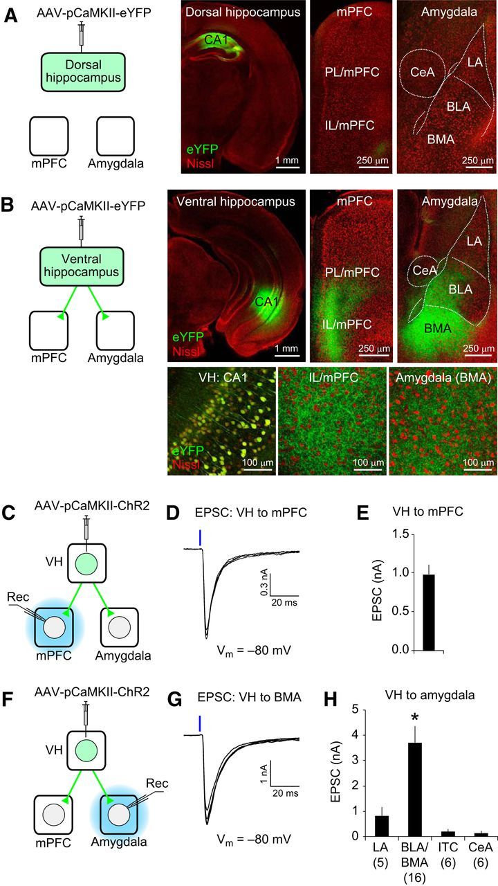
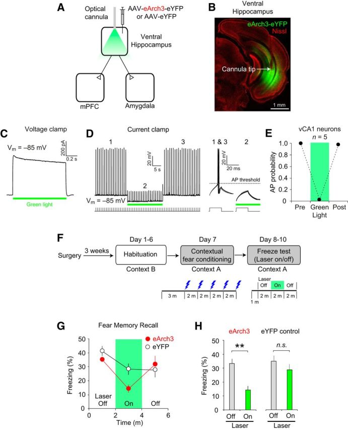
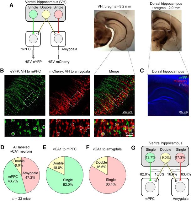
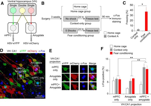
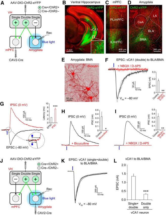
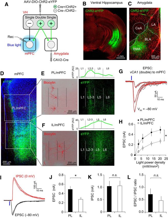
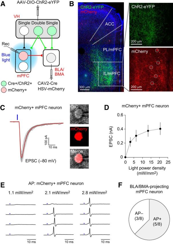
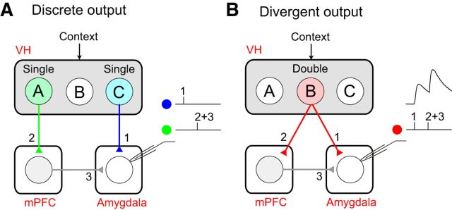
The acquisition and retrieval of contextual fear memory requires coordinated neural activity in the hippocampus, medial prefrontal cortex (mPFC), and amygdala. The contextual information encoded in the hippocampus is conveyed to the mPFC and amygdala for contextual fear conditioning. Previous studies have suggested that a CA1 neuronal population in the ventral hippocampus (VH) projects to both the mPFC and amygdala and is recruited in context-dependent control of conditioned fear. However, how double-projecting ventral CA1 hippocampal (vCA1) neurons modulate the activity of the mPFC and amygdala at the synaptic level has not been determined previously. Here, we show that the optogenetic silencing of the VH prevented the recall of contextual fear memory in mice, indicating its role in contextual fear expression. In dual retrograde viral tracing and c-Fos immunostaining experiments, we found that a proportion of vCA1 neurons projected to both the mPFC and amygdala and were recruited preferentially during context exposure, suggesting their role in encoding context representations. Moreover, optogenetic stimulation of axon collaterals of double-projecting vCA1 neurons induced monosynaptic excitatory responses in both the mPFC and basal amygdala, indicating that they could convey contextual information through the VH-mPFC and VH-amygdala pathways. The activation of double-projecting vCA1 neurons also induced action potential firings in the mPFC neurons that project to the amygdala, suggesting that they can also activate the VH-mPFC-amygdala pathway. With these synaptic mechanisms, double-projecting vCA1 neurons could induce synchronized neural activity in the mPFC and amygdala and convey contextual information efficiently to the basal amygdala for contextual fear conditioning.SIGNIFICANCE STATEMENT This work demonstrates that ventral CA1 hippocampal (vCA1) neurons projecting to both the medial prefrontal cortex (mPFC) and amygdala are activated preferentially when contextual information is processed in the ventral hippocampus, which is required for contextual fear expression. Our electrophysiological experiments reveal that the activation of double-projecting vCA1 neurons induces excitatory synaptic activity in both the mPFC and amygdala. These results suggest that double-projecting vCA1 neurons could contribute to contextual fear responses by inducing synchronized activity in the mPFC and amygdala and conveying contextual information to the basal amygdala more efficiently than vCA1 neurons projecting to either the mPFC or amygdala alone. These findings provide important insights into the mechanisms of the acquisition and retrieval of contextual fear memory.

Keywords: amygdala; fear conditioning; hippocampus; learning and memory; mPFC.


Figures

Figure 1.
Figure 1.
The VH projects directly to the mPFC and basal amygdala. A, Left, AAV-pCaMKII-eYFP was injected into the CA1 area of the DH. eYFP was expressed in dorsal CA1 neurons under the control of the CaMKII promoter (pCaMKII). Right, eYFP expression in the DH, mPFC, and amygdala (eYFP in green; fluorescent Nissl stain in red). Few eYFP-expressing projections from the DH were detected in the mPFC and amygdala. LA, BLA, BMA, and CeA are the lateral, basolateral, basomedial and central nucleus of the amygdala, respectively. B, AAV-pCaMKII-eYFP was injected into the VH. Confocal microscopic images show eYFP expression (green fluorescence) in the VH and eYFP-labeled VH projections in the mPFC and amygdala at a low (top) or high magnification (bottom). Note eYFP-labeled cell bodies in the CA1 area of the VH and eYFP-labeled axons in the IL/mPFC and BMA in the magnified images (bottom). C, Experimental setup for D and E. AAV-pCaMKII-ChR2-eYFP was injected into the VH. ChR2-eYFP is expressed in vCA1 neurons under the control of the CaMKII promoter (pCaMKII). Blue light illumination was applied locally to the mPFC in brain slices (blue) to stimulate VH projections expressing ChR2 and induce the postsynaptic responses recorded in the mPFC (Rec). D, Representative traces of EPSCs in the VH–mPFC pathway. A blue vertical bar indicates blue light illumination (20 mW/mm2, 1 ms duration) onto ChR2-expressing VH projections in the mPFC. E, Average peak amplitude of EPSCs in the VH–mPFC pathway recorded as described in D. n = 6 cells. F, Experimental setup for G and H. AAV-pCaMKII-ChR2-eYFP was injected into the VH. Blue light illumination was applied locally to the amygdala in brain slices (blue) to stimulate ChR2-expressing VH projections and induce the postsynaptic responses recorded in the amygdala (Rec). G, Representative traces of EPSCs in the VH–amygdala pathway. EPSCs were induced as described in D and recorded in a BMA neuron. H, Quantification of the peak amplitudes of EPSCs evoked by stimulation of VH projections and recorded in different nuclei of the amygdala. The numbers in the parentheses are the number of neurons examined for each nucleus of the amygdala. *p < 0.05, BLA/BMA versus LA, intercalated cells (ITC), or CeA. Error bars indicate SEM.
Figure 2.
Figure 2.
Optogenetic silencing of the VH prevents the recall of contextual fear memory. A, AAV-eArch3-eYFP was injected and optical cannulae were implanted bilaterally into the VH to silence VH activity in vivo (eArch3 group, n = 5 mice). AAV-eYFP was injected into the VH in the eYFP control group (n = 7 mice). B, Microscopic image showing eArch-eYFP expression (green fluorescence) and the cannula implantation site (arrow). Red fluorescence indicates Nissl stain. C, Representative trace of eArch3-mediated currents induced by green light illumination (565 nm, 10 mW/mm2, green horizontal bar) and recorded in voltage-clamp mode at −85 mV in an eArch3-expressing vCA1 neuron in a brain slice. D, Left, Representative trace recorded in current-clamp mode showing the inhibition of action potentials (APs) by green light illumination (10 mW/mm2, green horizontal bar), which induced hyperpolarization in eArch3-expressing vCA1 neurons in brain slices. Right, Traces showing AP firings and their inhibition by green light illumination in a different time scale. The baseline membrane potential (Vm) was ∼−85 mV. APs were induced by square pulses of current injections (150 pA, 20 ms duration) at 1 Hz frequency as indicated below the traces. E, Quantification of AP firing probability in the presence or absence of green light illumination. AP firing was inhibited almost completely by green light illumination activating eArch3. n = 5 eArch3-expressing CA1 neurons. F, Experimental setup. After surgery, mice were habituated to handling and optical cable connection in context B for 6 d (days 1–6) and fear conditioned in context A on day 7. On days 8–10, freezing behavior was monitored in context A in the presence and absence of green light illumination onto the VH. G, Time course of freezing behavior in context A after contextual fear conditioning in the eArch3 (n = 5 mice, red closed circle) and eYFP groups (n = 7 mice, black open circles). After a 1 min acclimatization period and baseline recording of freezing behavior for 2 min, green light illumination was applied to the VH through bilateral optical cannulae for 2 min (green area within the graph). The freezing score was calculated as the percentage of time in which the mice remained immobile and the bout length was 2 s. For each mouse, freezing score (%) was calculated on each test day (days 8–10) and averaged for each time bin. H, Quantification of freezing behavior in context A. The recall of contextual fear memory was inhibited by optogenetic silencing of the VH in the eArch3 group, but not in the eYFP control group. Freezing scores on days 8–10 were averaged for each mouse. Freezing behavior was significantly reduced by green laser illumination in the eArch3 group (**p < 0.01, laser on vs laser off), whereas it was not affected by laser illumination in the eYFP control group (n.s., not significant, laser on vs laser off). Error bars indicate SEM.
Figure 3.
Figure 3.
Some ventral CA1 hippocampal neurons project to both the mPFC and amygdala. A, Left, Retrograde viral tracers HSV-eYFP and HSV-mCherry were injected into the mPFC and basal amygdala (BLA and BMA), respectively. Right, Microscopic images of the coronal brain sections containing the DH and VH. Dotted squares indicate the areas magnified in B and C. B, Microscopic images showing vCA1 neurons projecting to the mPFC (eYFP-positive, green fluorescence, left) and to the amygdala (mCherry-positive, red fluorescence, middle). These images are overlaid in the right panels and show double-projecting vCA1 neurons expressing both eYFP and mCherry (yellow). The pyramidal cell layer of the vCA1 area (dotted squares) in each fluorescence channel is shown at a higher magnification in the bottom and double-projecting vCA1 neurons are indicated with circles. C, Microscopic image of the DH from a mouse receiving injections of HSV-eYFP and HSV-mCherry into the mPFC and amygdala, as described in A. Note the lack of eYFP- and mCherry-positive neurons in the DH. Blue fluorescence represents DAPI staining. D, Pie chart showing the average proportion of vCA1 neurons projecting to the mPFC and/or amygdala of all labeled vCA1 neurons (n = 22 mice). E, Pie chart showing the average proportion of double-projecting vCA1 neurons of all CA1 neurons projecting to the mPFC (n = 22 mice). F, Pie chart showing the average proportion of double-projecting vCA1 neurons of all CA1 neurons projecting to the basal amygdala (n = 22 mice). G, Diagram summarizing the results in DF.
Figure 4.
Figure 4.
Double-projecting vCA1 neurons are activated preferentially during context exposure and the recall of contextual fear memory. A, Retrograde HSV-eYFP and HSV-mCherry were injected into the mPFC and basal amygdala to label vCA1 neurons projecting to these areas. B, On the training day, mice in the fear-conditioning group received 5 electric foot shocks (0.5 mA, 2 s duration) in a novel context A, whereas the mice in the context-only group were exposed to the context without an aversive stimulus. After 24 h, these mice were tested for freezing behavior in context A. Ninety minutes after the behavioral test, brain tissues from these mice were fixed for c-Fos immunostaining. Mice in the home cage control group were left in home cages until brain fixation. n = 6 mice per group. C, Quantification of freezing behavior in context A on the test day in the context-only and fear-conditioning groups. *p < 0.05. D, Microscopic image of vCA1 pyramidal neurons. vCA1 neurons projecting to the mPFC and amygdala were labeled with eYFP (green) and mCherry (red), respectively. Double-projecting vCA1 neurons were labeled with both eYFP and mCherry (mPFC + amygdala, yellow). Blue fluorescence indicates c-Fos expression in vCA1 neurons. Squares indicate the areas magnified in E. E, Microscopic images showing examples of c-Fos-positive vCA1 neurons projecting to the mPFC and/or amygdala in different fluorescent channels. F, Quantification of the proportion of c-Fos-positive vCA1 neurons projecting to the mPFC and/or amygdala for each behavioral group. When the mice were exposed to the context or recalled contextual fear memory, a greater proportion of double-projecting vCA1 neurons expressed c-Fos compared with vCA1 neurons projecting to either the mPFC or amygdala alone (**p < 0.01). Error bars indicate SEM.
Figure 5.
Figure 5.
Double-projecting vCA1 neurons make excitatory synapses onto BLA/BMA neurons. A, Experimental setup for BI. Retrograde CAV2-Cre was injected into the mPFC, and AAV-DIO-ChR2-eYFP was injected into the CA1 area of the VH, resulting in the expression of Cre recombinase (Cre+) and ChR2-eYFP (ChR2+) in vCA1 neurons projecting to the mPFC. Local blue light illumination onto the amygdala activated the axon collaterals of double-projecting vCA1 neurons, inducing postsynaptic responses in the amygdala (Rec). vCA1 neurons projecting to the amygdala, but not to the mPFC, did not express Cre or ChR2 (Cre−/ChR2−) and their axons were not activated by local photostimulation. BD, Confocal microscopic images of the VH, mPFC, and amygdala taken 4 weeks after viral injection surgery, as described in A. Green fluorescence represents eYFP and red fluorescence represents Nissl stain. B, Green fluorescence in the VH indicates eYFP-labeled vCA1 pyramidal neurons projecting to the mPFC (inset is a magnified image of vCA1 neurons). C, Green fluorescence in the mPFC indicates the eYFP-labeled axons of mPFC-projecting vCA1 neurons. D, Green fluorescence in the amygdala indicates axons from vCA1 neurons projecting to both the mPFC and basal amygdala (BLA/BMA). E, Representative image showing three principal neurons in the BMA loaded with biocytin (red fluorescence) during the whole-cell patch-clamp recording. These BMA neurons were labeled with streptavidin–Alexa Fluor 568 after recording. F, Left, Representative traces of EPSCs induced by photostimulation of the axons of double-projecting vCA1 neurons and recorded in a BMA neuron. EPSCs were evoked by photostimulation (470 nm blue LED, 20 mW/mm2, 1 ms duration, 0.05 Hz stimulation frequency, blue vertical bar) applied locally onto the amygdala in brain slices and recorded at −80 mV in voltage-clamp mode. The black trace is the average of individual EPSCs (gray traces) under control conditions. EPSCs were completely blocked by the application of NBQX (10 μm) and D-AP5 (50 μm), shown as a red trace, which is the average of individual EPSCs (pink traces). Right, Quantification of EPSC inhibition by NBQX and D-AP5 (**p < 0.01, n = 7 cells). G, Representative traces of the EPSC (black trace) and IPSC (red trace) recorded in a BMA neuron. Synaptic responses were induced by photostimulation of double-projecting vCA1 neurons (blue vertical bars), as described in F. EPSCs and IPSCs were recorded at −80 and 0 mV, respectively, in voltage-clamp mode in the same BMA neuron. These traces are the average of three individual traces of EPSCs or IPSCs. The initial portion of the postsynaptic responses is magnified in a dotted circle. Inset, Graph showing the average synaptic delay of EPSCs and IPSCs. The onset of IPSCs was more delayed than EPSCs (***p < 0.001, n = 12 cells). H, Representative traces of IPSCs induced and recorded as described in G. The black trace is IPSCs recorded under control conditions. IPSCs were completely blocked by the application of bicuculine (30 μm, red trace). Inset, Quantification of EPSC inhibition by bicuculline (*p < 0.05, n = 6 cells). I, Representative traces of IPSCs induced and recorded as described in G. The black trace is IPSCs recorded under control conditions. IPSCs were completely blocked by the application of NBQX (10 μm) + D-AP5 (50 μm) (red trace). Inset, Quantification of EPSC inhibition by NBQX (10 μm) + D-AP5 (**p < 0.01, n = 6 cells). J, Experimental setup for K. Retrograde CAV2-Cre was injected into the basal amygdala (BLA/BMA) and AAV-DIO-ChR2-eYFP was injected into the VH CA1 area, resulting in the expression of Cre recombinase (Cre+) and ChR2-eYFP (ChR2+) in vCA1 neurons projecting to the basal amygdala. Local blue light illumination onto the amygdala activated the presynaptic axons of vCA1 neurons projecting to the basal amygdala alone (single-projecting neurons), as well as vCA1 neurons projecting to both the mPFC and amygdala (double-projecting neurons), which induced postsynaptic responses in the basal amygdala (Rec). K, Representative traces of EPSCs induced by photostimulation of axons of amygdala-projecting vCA1 neurons (single and double projecting). EPSCs were evoked by photostimulation (470 nm blue LED, 20 mW/mm2, 1 ms duration, blue vertical bar) applied locally to the amygdala and were recorded in a BMA neuron at −80 mV in voltage-clamp mode, as described in F. Note the different vertical scale bars in F and K. L, Quantification of the peak amplitudes of EPSCs recorded in the basal amygdala and induced by photostimulation, as described in F and K. Photostimulations of the same light intensity and duration (20 mW/mm2, 1 ms duration) were applied to activate the axons of both single- and double-projecting vCA1 neurons, as described in J and K, or stimulate presynaptic inputs only from double-projecting vCA1 neurons, as described in A and F. EPSCs induced by optogenetic stimulation of both single-and double-projecting vCA1 neurons were significantly larger than EPSCs induced by activation of double-projecting vCA1 neurons alone (***p < 0.001). n = 23 cells for single/double-projecting vCA1 axons and n = 24 cells for double-projecting vCA1 axons. Error bars indicate SEM.
Figure 6.
Figure 6.
Double-projecting vCA1 neurons form functional synapses onto mPFC neurons. A, Experimental setup. Retrograde CAV2-Cre was injected into the basal amygdala (BLA/BMA) and AAV-DIO-ChR2-eYFP was injected into the VH, inducing the expression of Cre recombinase and ChR2-eYFP (Cre+/ChR2+) in vCA1 neurons projecting to the basal amygdala. Local blue light illumination onto the mPFC activated the ChR2-expressing axons of double-projecting vCA1 neurons, inducing postsynaptic responses in the mPFC (Rec). vCA1 neurons projecting to the mPFC, but not to the amygdala, did not express Cre or ChR2 (Cre−/ChR2−) and their axons were not activated by local photostimulation. B, C, Confocal microscopic images of the VH (B) and amygdala (C) taken 4 weeks after viral injection surgery, as described in A. B, Green fluorescence in the VH indicates CA1 pyramidal neurons projecting to the basal amygdala. C, Green fluorescence in the amygdala indicates axon fibers of amygdala-projecting vCA1 neurons. Red fluorescence indicates Nissl staining. D, Microscopic image showing eYFP-labeled axons (green) of double-projecting vCA1 neurons in the mPFC. Parts of the PL and IL divisions of the mPFC are marked with square dotted lines and are shown in E and F, respectively. Blue fluorescence indicates DAPI staining. E, Magnified images of the PL/mPFC in D. Left, Three pyramidal neurons in layers 2–3 of the PL/mPFC were labeled with biocytin/streptavidin–Alexa Fluor 568 (red) after electrophysiological recording. Right, Same area of the PL/mPFC as in the left panel showing the axonal fibers of double-projecting vCA1 neurons (green). These projections were found predominantly in layers 2–3 (L2–L3) of the PL/mPFC. The borders between the cortical layers are indicated as dotted vertical lines. The graph above the image indicates the relative intensity of eYFP (green, arbitrary unit, AU) in the PL/mPFC. F, Magnified images of the IL/mPFC in D. Left, Two pyramidal neurons in L5 of the IL/mPFC were labeled with biocytin/streptavidin–Alexa Fluor 568 (red) after electrophysiological recording. Right, Same area of the IL/mPFC as in the left panel showing the axonal fibers of double-projecting vCA1 neurons (green). These projections were found in L2–L3 and L5–L6) of the IL/mPFC. The borders between the cortical layers are indicated as dotted vertical lines. The graph above the image indicates the relative intensity of eYFP in the IL/mPFC. G, Representative traces of EPSCs recorded in an mPFC neuron. EPSCs were evoked by pulses of photostimulation (470 nm blue LED, 1 ms duration, blue vertical bar) of the ChR2-expressing axons of double-projecting vCA1 neurons and were recorded in the mPFC neuron at −80 mV in voltage-clamp mode. Three groups of EPSCs were evoked by photostimulation with different light intensities (2.8, 6.4, and 13.4 mW/mm2). The red traces are the averages of individual EPSCs (gray traces). H, Graph showing the average amplitudes of EPSCs induced and recorded as described in G, which are plotted versus photostimulation intensity. The peak amplitudes of EPSCs in the PL/mPFC (filled circles) were significantly larger than in the IL/mPFC (open circles). *p < 0.05. n = 10 and 11 neurons (4 mice) for PL and IL, respectively. I, Representative traces of EPSCs (black traces) and IPSCs (red traces) recorded in an mPFC neuron. The EPSCs and IPSCs were induced as described in G and recorded at −80 and 0 mV, respectively, in voltage-clamp mode in the same mPFC neuron. Note a more delayed onset of IPSC than EPSC after photostimulation (20 mW/mm2, blue vertical line). JL, Quantification of the peak amplitudes of the EPSC (J) and IPSC (K) and the EPSC/IPSC ratio (L) in postsynaptic PL/mPFC and IL/mPFC neurons. In each mPFC neuron, both the EPSCs and IPSCs were evoked by photostimulation of the same intensity (20 mW/mm2). The average EPSC was significantly larger in the PL/mPFC than in the IL/mPFC (J, *p < 0.05). There was no significant difference in IPSC amplitude (K; n.s., not significant) or the EPSC/IPSC ratio (L) between the PL and IL (n.s., not significant). n = 8–10 cells (4 mice) for the PL and n = 8–11 cells (4 mice) for the IL. Error bars indicate SEM.
Figure 7.
Figure 7.
Double-projecting vCA1 neurons activate amygdala-projecting mPFC neurons of the VH–mPFC–amygdala circuit. A, Experimental setup. Retrograde CAV2-Cre was injected into the basal amygdala (BLA/BMA) and AAV-DIO-ChR2-eYFP was injected into the VH to induce ChR2-eYFP expression in the vCA1 neurons projecting to the basal amygdala. Another retrograde virus, HSV-mCherry, was also injected into the basal amygdala to label the BLA/BMA-projecting mPFC neurons with mCherry. Local blue light illumination onto the mPFC activated the axons of double-projecting CA1 neurons expressing ChR2-eYFP, which induced the postsynaptic responses recorded in BLA/BMA-projecting mPFC neurons (Rec). B, Left, Microscopic image showing the eYFP-labeled axons of double-projecting vCA1 neurons in the mPFC (green). Blue fluorescence represents DAPI staining. A portion of the IL/mPFC (a dotted square) is magnified in the right panels. Top right, eYFP-labeled axons of double-projecting vCA1 neurons in the IL/mPFC (green). Bottom right, mCherry-labeled IL/mPFC neurons projecting to the basal amygdala (red, dotted circles). ACC, Anterior cingulate cortex. C, Left, Representative traces of EPSCs recorded in an amygdala-projecting mPFC neuron. EPSCs were induced by photostimulation (470 nm blue LED, 1 ms duration, blue vertical bar) of the ChR2-expressing axons of double-projecting vCA1 neurons and recorded in mCherry-labeled mPFC neurons at −80 mV in voltage-clamp mode. The red trace is the average of the individual EPSCs (gray traces). Right, Microscopic images of the mCherry-labeled recorded neuron confirming that the mPFC neuron projects to the basal amygdala. DIC, Differential interference contrast microscopic image. D, Graph showing the average peak amplitudes of EPSCs recorded, as described in C, which are plotted versus photostimulation intensity. n = 10 neurons from 2 mice. Error bars indicate SEM. E, Representative traces of action potential (AP) firings recorded in an mCherry-labeled mPFC neuron in cell-attached mode. The intensity of photostimulation (blue bars) is indicated above each column. F, Photostimulation of double-projecting vCA1 neurons induced AP firings in 5 of 8 amygdala-projecting mPFC neurons examined (AP+: 62.5%).
Figure 8.
Figure 8.
Double-projecting vCA1 neurons can induce synchronized neural activity in the mPFC and amygdala and activate the basal amygdala more efficiently than single-projecting CA1 neurons. A, Discrete output of the VH. Single-projecting vCA1 neurons A (green) and C (blue) encode specific contextual information. Neuron A projects only to the mPFC (pathway 2) and neuron C projects only to the amygdala (pathway 1). It is less likely that VH activity with discrete output can induce synchronized activity in the mPFC and amygdala unless neurons A and C are activated simultaneously. Neurons A and C induce excitatory postsynaptic responses in the basal amygdala through the indirect VH–mPFC–amygdala pathway (pathways 2 + 3) and the direct VH–amygdala pathway (pathway 1), respectively. Temporally separated activation of neurons A and C induces less efficiently temporal summation of the excitatory postsynaptic responses in the basal amygdala. B, Divergent output of the VH. vCA1 neuron B (red) encodes specific contextual information and projects to both the mPFC and amygdala. The activation of neuron B induces excitatory postsynaptic responses in both the mPFC (pathway 2) and amygdala (pathway 1) simultaneously through axon collaterals (Figs. 5 and 6), which facilitated the synchronized neural activity in these areas that is implicated in conditioned fear (Lesting et al., 2011). Moreover, double-projecting neuron B can also activate the basal amygdala more efficiently than single-projecting vCA1 neurons. The activation of neuron B induces action potential firings in mPFC neurons projecting to the basal amygdala (Figs. 7), resulting in additional excitatory postsynaptic responses in the amygdala through the indirect VH–mPFC–amygdala pathway (pathways 2 + 3). The simultaneous activation of both the direct VH–amygdala pathway and the indirect VH–mPFC–amygdala pathway by double-projecting neuron B facilitates the temporal summation of excitatory postsynaptic responses in the basal amygdala, which may contribute to the induction of long-term synaptic potentiation in the VH–amygdala pathway during the acquisition of contextual fear memory.

References

    1. Adhikari A, Lerner TN, Finkelstein J, Pak S, Jennings JH, Davidson TJ, Ferenczi E, Gunaydin LA, Mirzabekov JJ, Ye L, Kim SY, Lei A, Deisseroth K (2015) Basomedial amygdala mediates top-down control of anxiety and fear. Nature 527:179–185. 10.1038/nature15698 - DOI - PMC - PubMed
    1. Atasoy D, Aponte Y, Su HH, Sternson SM (2008) A FLEX switch targets Channelrhodopsin-2 to multiple cell types for imaging and long-range circuit mapping. J Neurosci 28:7025–7030. 10.1523/JNEUROSCI.1954-08.2008 - DOI - PMC - PubMed
    1. Balsam PD, Drew MR, Gallistel CR (2010) Time and associative learning. Comp Cogn Behav Rev 5:1–22. 10.3819/ccbr.2010.50001 - DOI - PMC - PubMed
    1. Bast T, Zhang WN, Feldon J (2001) The ventral hippocampus and fear conditioning in rats. Different anterograde amnesias of fear after tetrodotoxin inactivation and infusion of the GABA(A) agonist muscimol. Exp Brain Res 139:39–52. 10.1007/s002210100746 - DOI - PubMed
    1. Beyeler A, Namburi P, Glober GF, Simonnet C, Calhoon GG, Conyers GF, Luck R, Wildes CP, Tye KM (2016) Divergent routing of positive and negative information from the amygdala during memory retrieval. Neuron 90:348–361. 10.1016/j.neuron.2016.03.004 - DOI - PMC - PubMed

Publication types

MeSH terms