. 2017 Apr 6;7(1):665.
doi: 10.1038/s41598-017-00706-7.

Discovery and characterization of verinurad, a potent and specific inhibitor of URAT1 for the treatment of hyperuricemia and gout

Affiliations

Discovery and characterization of verinurad, a potent and specific inhibitor of URAT1 for the treatment of hyperuricemia and gout

Philip K Tan et al. Sci Rep. .

Abstract

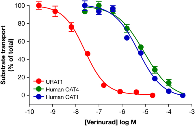
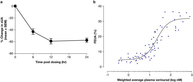
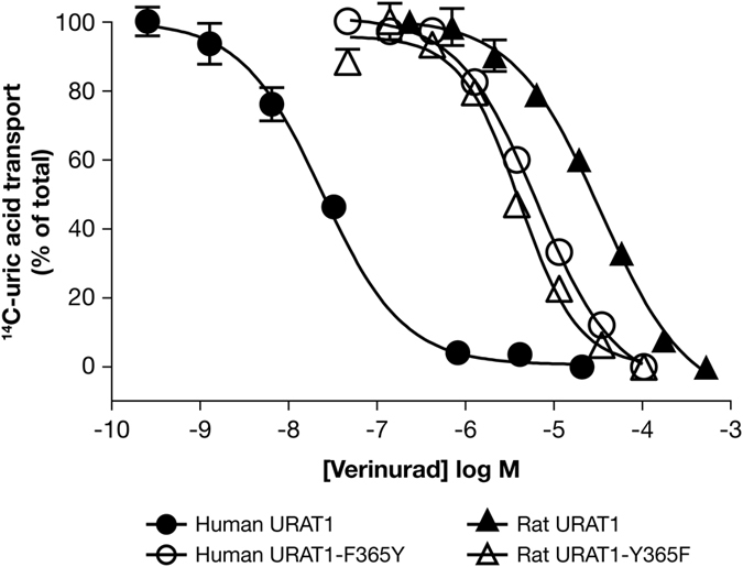
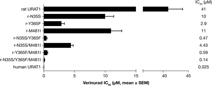
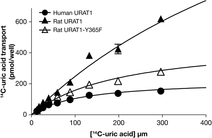
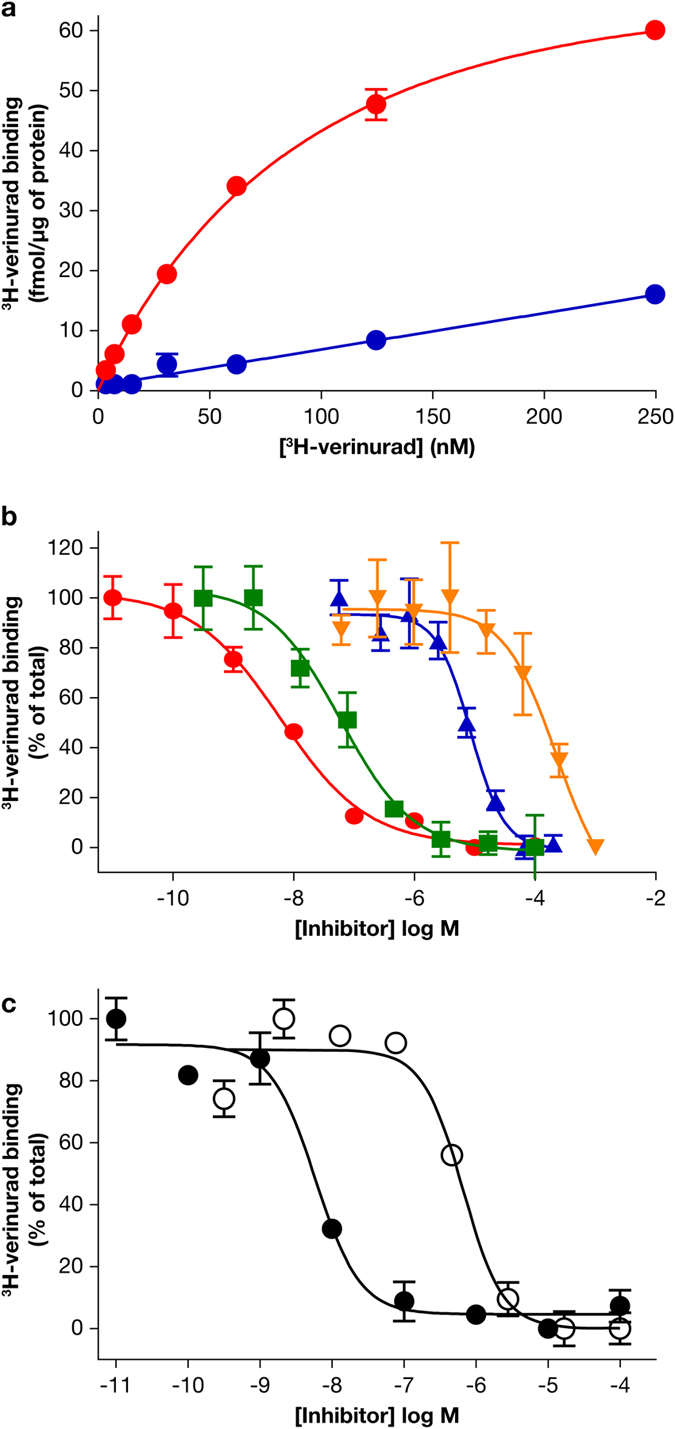
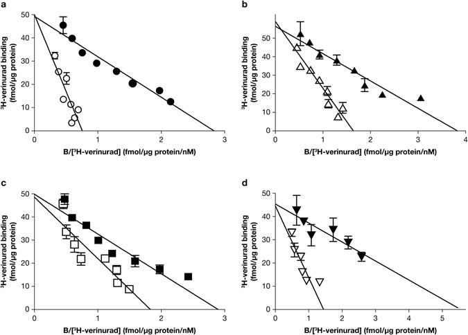
Gout is caused by elevated serum urate levels, which can be treated using inhibitors of the uric acid transporter, URAT1. Here, we characterize verinurad (RDEA3170), which is currently under evaluation for gout therapy. Verinurad specifically inhibits URAT1 with a potency of 25 nM. High affinity inhibition of uric acid transport requires URAT1 residues Cys-32, Ser-35, Phe-365 and Ile-481. Unlike other available uricosuric agents, the requirement for Cys-32 is unique to verinurad. Two of these residues, Ser-35 and Phe-365, are also important for urate transport kinetics. A URAT1 binding assay using radiolabeled verinurad revealed that distinct URAT1 inhibitors benzbromarone, sulfinpyrazone and probenecid all inhibit verinurad binding via a competitive mechanism. However, mutations made within the predicted transporter substrate channel differentially altered the potency for individual URAT1 inhibitors. Overall, our results suggest that URAT1 inhibitors bind to a common site in the core of the transporter and sterically hinder the transit of uric acid through the substrate channel, albeit with vastly different potencies and with differential interactions with specific URAT1 amino acids.


Conflict of interest statement

The authors declare that they have no competing interests.

Figures

Figure 1
Figure 1
Verinurad is highly potent and specific for human URAT1. Dose-responses for verinurad against the transport activity of human URAT1 (red), human OAT4 (green) and human OAT1 (blue). Cells expressing URAT1 were incubated with 14C-uric acid, and cells expressing OAT4 or OAT1 were incubated with carboxyfluorescein, in the presence of different amounts of verinurad. Data are from one representative experiment presented as the mean ± SEM of samples measured in triplicate.
Figure 2
Figure 2
Verinurad lowers serum uric acid (sUA) by increasing fractional excretion of uric acid (FEUA) in humans. (a) A single 40 mg dose of verinurad lowered the baseline sUA levels by up to 60% for a sustained time period. (b) Verinurad increased the FEUA in a dose-dependent manner, with a half-maximal effective plasma concentration 22 nM. Experiments were performed in healthy human volunteers.
Figure 3
Figure 3
Phe-365 of human URAT1 is important for affinity for verinurad. Dose-response curves of verinurad against human URAT1 (solid circles), human URAT1-F365Y (open circles), rat URAT1 (solid triangles) and rat URAT1-Y365F (open triangles). The experiment was performed as described in Fig. 1. The potency of verinurad for each construct can be found in Table 2. Data are from one representative experiment presented as the mean ± SEM of samples measured in triplicate.
Figure 4
Figure 4
Human URAT1 residues 35, 365 and 481 cooperate to increase affinity for verinurad. Potency of verinurad against rat URAT1, individual or combination chimeric point mutants with human URAT1 residues at positions 35, 365 and 481, and human URAT1. Dose-response experiments were performed as in Fig. 3, and results are the mean ± SEM from at least three experiments.
Figure 5
Figure 5
Human URAT1 Ser-35 and Phe-365 cooperate to enhance urate affinity. Transport kinetics of human URAT1 (filled circles), rat URAT1 (filled triangles) and the double point mutant chimera rat URAT1 carrying human URAT1 residues at 35 and 365 (r-N35S/F365Y, open triangles). Data are from one representative experiment presented as the mean ± SEM of samples measured in triplicate. The half maximal transport velocity (K m) for each construct is presented in Table 3.
Figure 6
Figure 6
Each URAT1 inhibitor has a distinct profile of potencies for URAT1 mutants. Potencies of verinurad (a), benzbromarone (b), sulfinpyrazone (c) and probenecid (d) against human URAT1 (hURAT1) and hURAT1 with “binding site” point mutants F241Y, F449Y and R477K. Dose-response experiments were performed as in Fig. 3, and results are the mean ± SEM from at least three experiments.
Figure 7
Figure 7
A novel URAT1 binding assay replicates the results of inhibition of URAT1 activity. (a) 3H-verinurad bound specifically to human URAT1. 3H-verinurad bound to membranes from cells transfected with human URAT1 in a specific and saturable manner (red) but not to membranes from cells transfected with empty vector (blue). (b) 3H-verinurad binding to human URAT1 was blocked by URAT1 inhibitors in a dose-dependent manner. 10 nM of 3H-verinurad was incubated with human URAT1 membranes and different concentrations of unlabeled verinurad (red), benzbromarone (green), sulfinpyrazone (blue) and probenecid (orange). (c) 3H-verinurad bound to membranes from cells transfected with human URAT1-F449Y, which was blocked in a dose-dependent manner by unlabeled verinurad (filled circles) and by benzbromarone (open circles). Compared to human URAT1, h-F449Y has the same affinity for verinurad but an 11-fold lower affinity for benzbromarone, mimicking the results for the activity assay (Fig. 6).
Figure 8
Figure 8
Inhibitors bind to the same binding site on URAT1. Binding of 3H-verinurad to human URAT1 in the absence of inhibitors (filled symbols) or in the presence of 20 nM unlabeled verinurad (a) (open circles), 50 nM benzbromarone (b) (open upward triangles), 25 µM sulfinpyrazone (c) (open squares) or 200 µM probenecid (d) (open downward triangles). Membranes containing human URAT1 were incubated with different concentrations of 3H-verinurad without or with inhibitors. Specifically bound 3H-verinurad is shown by Eadie-Hofstee linearization. B = bound verinurad.

References

    1. Choi HK, Mount DB, Reginato AM. Pathogenesis of gout. Ann. Intern. Med. 2005;143:499–516. doi: 10.7326/0003-4819-143-7-200510040-00009. - DOI - PubMed
    1. Neogi T. Clinical practice. Gout. N Engl J Med. 2011;364:443–452. doi: 10.1056/NEJMcp1001124. - DOI - PubMed
    1. Kratzer JT, et al. Evolutionary history and metabolic insights of ancient mammalian uricases. Proc. Natl. Acad. Sci. USA. 2014;111:3763–3768. doi: 10.1073/pnas.1320393111. - DOI - PMC - PubMed
    1. Tan PK, Farrar JE, Gaucher EA, Miner JN. Coevolution of URAT1 and uricase during primate evolution: implications for serum urate homeostasis and gout. Mol. Biol. Evol. 2016;33:2193–200. doi: 10.1093/molbev/msw116. - DOI - PMC - PubMed
    1. Levinson DJ, Sorensen LB. Renal handling of uric acid in normal and gouty subject: evidence for a 4-component system. Ann. Rheum. Dis. 1980;39:173–179. doi: 10.1136/ard.39.2.173. - DOI - PMC - PubMed

MeSH terms