. 2017 Apr 6;7(1):700.
doi: 10.1038/s41598-017-00662-2.

Delivery of ENaC siRNA to epithelial cells mediated by a targeted nanocomplex: a therapeutic strategy for cystic fibrosis

Affiliations

Delivery of ENaC siRNA to epithelial cells mediated by a targeted nanocomplex: a therapeutic strategy for cystic fibrosis

Maria D I Manunta et al. Sci Rep. .

Abstract

The inhibition of ENaC may have therapeutic potential in CF airways by reducing sodium hyperabsorption, restoring lung epithelial surface fluid levels, airway hydration and mucociliary function. The challenge has been to deliver siRNA to the lung with sufficient efficacy for a sustained therapeutic effect. We have developed a self-assembling nanocomplex formulation for siRNA delivery to the airways that consists of a liposome (DOTMA/DOPE; L), an epithelial targeting peptide (P) and siRNA (R). LPR formulations were assessed for their ability to silence expression of the transcript of the gene encoding the α-subunit of the sodium channel ENaC in cell lines and primary epithelial cells, in submerged cultures or grown in air-liquid interface conditions. LPRs, containing 50 nM or 100 nM siRNA, showed high levels of silencing, particularly in primary airway epithelial cells. When nebulised these nanocomplexes still retained their biophysical properties and transfection efficiencies. The silencing ability was determined at protein level by confocal microscopy and western blotting. In vivo data demonstrated that these nanoparticles had the ability to silence expression of the α-ENaC subunit gene. In conclusion, these findings show that LPRs can modulate the activity of ENaC and this approach might be promising as co-adjuvant therapy for cystic fibrosis.


Conflict of interest statement

The authors declare that they have no competing interests.

Figures
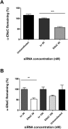
Figure 1
Figure 1
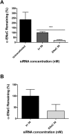
Silencing efficiency of cell lines transfected with LPRs containing peptide E. (A) In independent experiments, using 50 nM siRNA, the level of expression of α-ENaC gene in 16HBE14o- was of 57.2% in respect to GAPDH housekeeping gene (n = 7). Statistically significant difference was determined as ***P ≤ 0.001. (B) In A549 cells the remaining level of ENaC, expressed as percentage of non-targeting/irrelevant siRNA, was 53.4% at 50 nM, with a statistical difference of **P ≤ 0.01, and 67.5% at 100 nM. The results show the means ± S.E.M. of triplicate experiments.
Figure 2
Figure 2
Knockdown of α-ENaC in primary cells transfected with peptide E nanoparticles. (A) The α-ENaC expression in NHBE submerged cultures (n = 5) treated with 50 nM siRNA was 21.5% relative to GAPDH housekeeping gene. The difference was statistically significant (*** P ≤ 0.001). (B) Air-liquid interface cultures of NHBE (n = 3) treated with LPRs containing 50 nM siRNA showed a remaining expression of 33.8% giving therefore a silencing of 66.2% relative to GAPDH housekeeping gene. The remaining expression of the α-ENaC gene is expressed as percentage relative to non-targeted/irrelevant siRNA-treated cells.
Figure 3
Figure 3
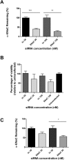
Primary CF epithelial cells with LPRs containing either 50 nM or 100 nM siRNA and peptide Y. (A) Percentage of expression of the α-ENaC mRNA after siRNA treatment in primary CF cells in submerged culture. The expression of α-ENaC was 40.5% at 50 nM and 27.2% at 100 nM, respectively. The difference was statistically significant (*** P ≤ 0.001) at 50 nM, whereas the significance (**P ≤ 0.01), was lower at 100 nM. The results show the means ± S.E.M. of triplicate experiments. (B) Viability (percentage) of primary CF cells following transfections with LPRs formulated with peptide Y compared to untransfected cells. The α-ENaC-siRNA treated cells showed a viability of 72% at 50 nM siRNA concentration and 57% at 100 nM. Cell survival was assessed using an LDH assay measuring the light emitted at 610 nm. Results represent mean values of triplicate repeats ± S.E.M. (C) Percentage of remaining expression of the α-ENaC gene transcript in primary CFBE cells grown on ALI in transwell plates. The expression of the transcript of the α-ENaC gene was 71.8% at 30 nM and 56.7% at 50 nM. The difference was statistically significant only at 50 nM (*P ≤ 0.05). qRT-PCR analysis was performed in triplicate on individual samples. The mRNA levels of the α-ENaC gene are expressed as percentage of irrelevant/non-targeted control siRNA.
Figure 4
Figure 4
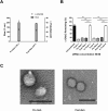
Nebulised LPRs. LPR particles were formulated at a 1:4:1 ratio of DOTMA/DOPE, peptide either Y or E and 100 μg of siRNA in 2 ml volume of H2O. (A) The particles sizes and the ζ potentials were assessed. The LPRs displayed sizes between 192 and 200 nm in diameter while the ζ potentials were between 23.6 and 25.6 mV. (B) The silencing capability of LPRs was detected after transfection of human airway epithelial (HAE) cells. The transfection ability of LPRs containing either peptide Y (n = 5) or peptide E (n = 6) and 50 nM siRNA was measured comparing the un-nebulised freshly made LPRs, the pre-nebulisation and post-nebulisation formulations relative to the freshly made non-targeting/irrelevant siRNA. Liposome-peptide Y- RNA nanoparticles freshly prepared or pre- and post-nebulisation displayed a silencing efficiency of 85.4%, 77.7% and 78.5%, respectively. Peptide E-LPRs showed similar levels of ENaC silencing 79.8% mediated by freshly prepared and pre-nebulisation, and 74.7% mediated by the nebulised LPRs. Statistically significant differences were determined as ***P ≤ 0.001. (C) LPRs pre- and post-nebulisation were also assessed by transmission electron microscopy (TEM). Size bars are 500 nm.
Figure 5
Figure 5
Confocal images of primary cells transfected with peptide E nanoparticles. Confocal images of NHBE cells grown in transwells and stained for α-ENaC (green), actin (red) and DAPI (blue) to label the nuclei; (A) untransfected control (B) non-targeting/irrelevant siRNA-treated and (C) α-ENaC siRNA-treated. The narrow panels on top and on the right side represent the cross-sectional view related to the indicated lines.
Figure 6
Figure 6
Western blotting of α-ENaC protein from A549 cells transfected with peptide E-LPRs. (A) Representative western blotting showing the α-ENaC major forms of 90 KDa and 65 KDa bands protein levels after treatment with LPR at both 50 nM and 100 nM siRNA, and GAPDH (36 KDa band). The lanes are indicated as following: untransfected control (U), treated with non-targeting/irrelevant siRNA (I) and α-ENaC siRNA-treated (E). (B) The densitometry results of 90 KDa α-ENaC protein from siRNA-treated cells extracted from the plasma membrane and from the cytosolic fraction, and expressed as percentage relative to the non-targeting/irrelevant siRNA-treated at a concentration of siRNA of 100 nM, showed that 50% of the subunit was still present at the cell surface, while 75% was still present in the cytosol.
Figure 7
Figure 7
In vivo delivery of LPRs containing 27 μg siRNA. (A) Following the LPRs administration, the knockdown in the expression of the α-ENaC gene mRNA in the lungs of C57BL6 female mice was measured by qRT-PCR. β actin was used as reference gene for mouse lung transcripts. The level of transcript expression of α-ENaC siRNA-treated group was compared with the control groups either untreated or non-targeting/irrelevant siRNA treated cohorts. Statistical analysis was performed using Student’s t-test. Statistical significance is represented by *P < 0.05. (B–D) Representative images of H&E stained lung sections from untreated control (B), non-targeting/irrelevant siRNA-treated (C) and α-ENaC siRNA-treated (D) mice show a mild peribronchial mononuclear cell infiltration in siRNA-treated animals (C,D) compared with untreated controls (B). Scale bars = 50 µm.

Similar articles

Cited by

References

    1. Riordan JR, et al. Identification of the cystic fibrosis gene: cloning and characterization of complementary DNA. Science. 1989;245:1066–1073. doi: 10.1126/science.2475911. - DOI - PubMed
    1. Buchwald M, Tsui LC, Riordan JR. The search for the cystic fibrosis gene. American Journal of Physiology. 1989;257:L47–52. - PubMed
    1. Mall MA, Galietta LJ. Targeting ion channels in cystic fibrosis. Journal of Cystic Fibrosis. 2015;14:561–570. doi: 10.1016/j.jcf.2015.06.002. - DOI - PubMed
    1. Canessa CM, et al. Amiloride-sensitive epithelial Na+ channel is made of three homologous subunits. Nature. 1994;367:463–467. doi: 10.1038/367463a0. - DOI - PubMed
    1. Rossier BC, Canessa CM, Schild L, Horisberger JD. Epithelial sodium channels. Current Opinion in Nephrology and Hypertension. 1994;3:487–496. doi: 10.1097/00041552-199409000-00003. - DOI - PubMed

Publication types

MeSH terms