. 2017 Feb 28;50(1):39-48.
doi: 10.1267/ahc.17003. Epub 2017 Feb 25.

Estrogen Accelerates Cell Proliferation through Estrogen Receptor α during Rat Liver Regeneration after Partial Hepatectomy

Affiliations

Estrogen Accelerates Cell Proliferation through Estrogen Receptor α during Rat Liver Regeneration after Partial Hepatectomy

Baatarsuren Batmunkh et al. Acta Histochem Cytochem. .

Abstract

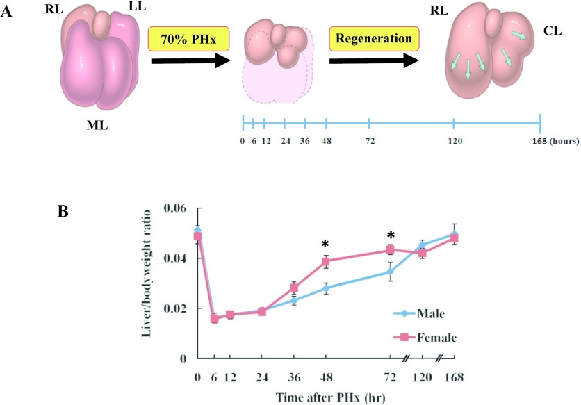
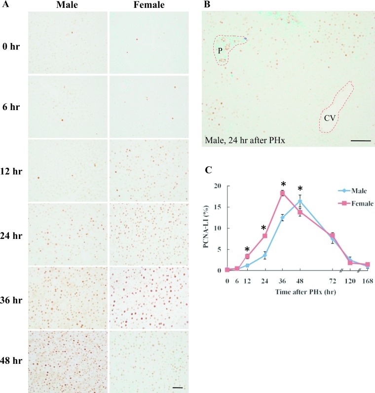
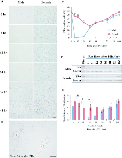
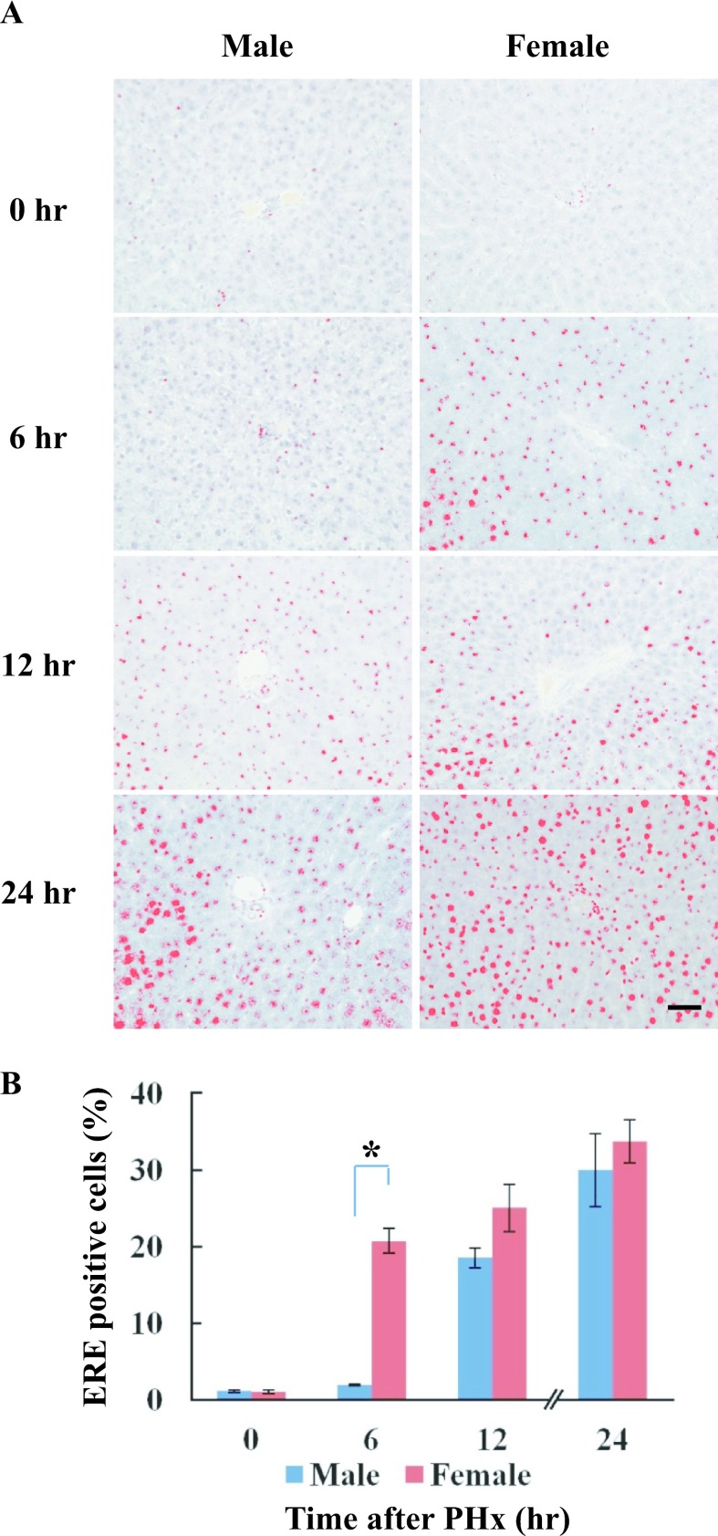
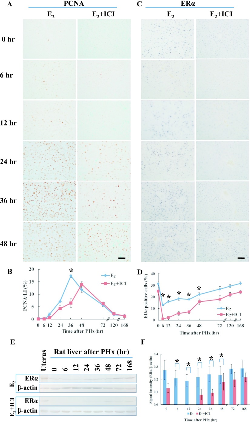
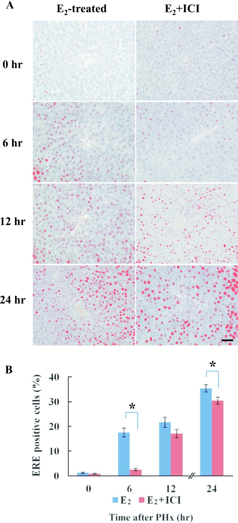
Although estrogen is implicated in the regulation of cell growth and differentiation in many organs, the exact mechanism for liver regeneration is not completely understood. We investigated the effect of estrogen on liver regeneration in male and female Wistar rats after 70% partial hepatectomy (PHx) and performed immunohistochemistry, western blotting and Southwestern histochemistry. 17β-estradiol (E2) and ICI 182,780 were injected into male rats on the day before PHx. The proliferating cell nuclear antigen (PCNA) labeling index reached a maximum at 48 hr after PHx in males, and at 36 hr in females and E2-treated male rats. Estrogen receptor α (ERα) was expressed in zones 1 and 2 in male rats, but was found in all zones in female rats. Interestingly, ERα was not detected at 6-12 hr after PHx but was found at 24-168 hr in male rats. However, ERα expression was found at all sampling time-points in female and E2-treated male rats. The activity of estrogen responsive element binding proteins was detected from 12 hr after PHx in male rats but was found from 6 hr in female and E2-treated male rats. ERα was co-expressed with PCNA during liver regeneration. These results indicate that estrogen may play an important role in liver regeneration through ERα.

Keywords: cell proliferation; estrogen; estrogen receptor α; liver regeneration; partial hepatectomy.


Figures

Fig. 1.
Fig. 1.
Model of liver regeneration and the liver weight/bodyweight ratio of male and female rats after PHx. A: Schema of 70% PHx in Wistar rats [13]. RL: right lobe, LL: left lobe, ML: median lobe, CL: caudate lobe. B: Liver weight/body weight ratio of male and female rats after PHx. Asterisks indicate statistically significant differences (*p < 0.05). Data represent the mean ± SE of 3–5 rats.
Fig. 2.
Fig. 2.
Immunohistochemical detection of PCNA in male and female rat liver after PHx. A: Liver tissue was collected from male and female rats at 0, 6, 12, 24, 36 and 48 hr after PHx. Paraffin-embedded rat liver sections were analyzed by immunohistochemistry. Magnification ×400. Bar = 50 μm. B: Zonal distribution of PCNA-positive cells at 24 hr in male rat liver after PHx. P: portal area, CV: central vein. Magnification ×200. Bar = 100 μm. C: PCNA-LI in male and female rats after PHx. The number of PCNA positive hepatocytes was counted at each time-point after PHx. Blue and red lines represent male and female, respectively. Asterisks indicate statistically significant differences (*p < 0.05). Data represent the mean ± SE of three independent experiments.
Fig. 3.
Fig. 3.
ERα expression in rat liver after PHx. A: Immunohistochemical detection of ERα in male and female rat liver at various time-points after PHx. Paraffin-embedded liver sections were analyzed by immunohistochemistry. Magnification ×400. Bar = 50 μm. B: Zone dependent ERα expression at 36 hr in male rat liver after PHx. P: portal area, CV: central vein. Magnification ×200. Bar = 100 μm. C: The number of ERα positive cells in the liver sections of male and female rats at various time-points after PHx. Blue and red lines represent male and female, respectively. D: Western blot analysis of ERα in male and female rats. ERα (66 kDa) and β-actin (42 kDa). E: Densitometry analysis of western blot. Asterisks indicate statistically significant differences (*p < 0.05). Data represent the mean ± SE of three independent experiments.
Fig. 4.
Fig. 4.
Localization of ERE binding proteins in male and female rats after PHx. Paraffin-embedded liver sections were used for detection of ERE binding proteins by Southwestern histochemistry at various time-points after PHx. A: The localization of ERE binding proteins was detected from 12 hr after PHx in male rats, but was found from 6 hr in female rats. Positive staining was processed using a DAB-image analyzer. B: The number of ERE-positive cells in the liver sections of male and female rats at various time-points after PHx. Blue and red columns represent male and female, respectively. Asterisk indicates statistically significant difference (*p < 0.05). Magnification ×400. Bar = 50 μm.
Fig. 5.
Fig. 5.
Double staining for PCNA and ERα in male and female rat liver after PHx. The paraffin-embedded sections were analyzed by immunohistochemistry. A: ERα-positive cells were stained brown (DAB), whereas PCNA positive cells were stained purple-blue (4-Cl-1-Naphtol). Boxed area is enlarged in B. Arrows indicate white (ERα), black (PCNA) and red (double staining), respectively. Magnification ×400. Scale Bar = 50 μm.
Fig. 6.
Fig. 6.
PCNA and ERα expression in E2 and E2+ICI treated male rat liver after PHx. A: Immunohistochemical detection of PCNA in E2 and E2+ICI treated male rat liver after PHx. B: PCNA-LI in E2 and E2+ICI treated rats after PHx. The number of PCNA-positive hepatocytes was counted at each time-point after PHx. Blue and red lines represent E2 or E2+ICI treated male rats, respectively. Data represent the mean ± SE of three independent experiments. C: Immunohistochemical detection of ERα in E2 and E2+ICI treated rats at various time-points after PHx. Magnification ×400. Bar = 50 μm. D: The number of ERα-positive cells in the liver sections of E2 and E2+ICI treated rats at various time-points after PHx. Blue and red lines represent E2 or E2+ICI treated male rats, respectively. E: Western blot analysis of ERα in E2 or E2+ICI treated male rats after PHx. ERα (66 kDa) and β-actin (42 kDa). F: Densitometry analysis of western blot. Asterisks indicate statistically significant differences (*p < 0.05). Data represent the mean ± SE of three independent experiments.
Fig. 7.
Fig. 7.
Localization of ERE binding proteins in E2 or E2+ICI treated male rats after PHx. A: The localization of ERE binding proteins was detected from 12 hr after PHx in E2+ICI treated rats, but was found from 6 hr in rats treated with E2. Positive staining was processed using a DAB-image analyzer. B: The number of ERE positive cells in the liver sections of E2 and E2+ICI treated male rats at various time-points after PHx. Blue and red columns represent E2 or E2+ICI treated male rats, respectively. Asterisks indicate statistically significant differences (*p < 0.05). Magnification ×400. Bar = 50 μm.

References

    1. Adams J. C. (1981) Heavy metal intensification of DAB-based HRP reaction product. J. Histochem. Cytochem. 29; 775. - PubMed
    1. An S., Soe K., Akamatsu M., Hishikawa Y. and Koji T. (2012) Accelerated proliferation of hepatocytes in rats with iron overload after partial hepatectomy. Histochem. Cell Biol. 138; 773–786. - PubMed
    1. Brady C. W. (2015) Liver disease in menopause. World J. Gastroenterol. 21; 7613–7620. - PMC - PubMed
    1. Choijookhuu N., Sato Y., Nishino T., Endo D., Hishikawa Y. and Koji T. (2012) Estrogen-dependent regulation of sodium/hydrogen exchanger-3 (NHE3) expression via estrogen receptor β in proximal colon of pregnant mice. Histochem. Cell Biol. 137; 575–587. - PubMed
    1. El Marzouk S., Gahattamaneni R., Joshi S. R. and Scovell W. M. (2008) The plasticity of estrogen receptor-DNA complexes: binding affinity and specificity of estrogen receptors to estrogen response element half-sites separated by variant spacers. J. Steroid Biochem. Mol. Biol. 110; 186–195. - PubMed