Review
. 2017 Mar 23:8:151.
doi: 10.3389/fphar.2017.00151. eCollection 2017.

ClC Channels and Transporters: Structure, Physiological Functions, and Implications in Human Chloride Channelopathies

Affiliations
Review

ClC Channels and Transporters: Structure, Physiological Functions, and Implications in Human Chloride Channelopathies

Diogo R Poroca et al. Front Pharmacol. .

Abstract

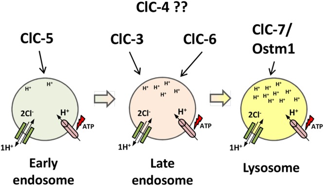
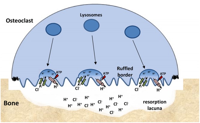
The discovery of ClC proteins at the beginning of the 1990s was important for the development of the Cl- transport research field. ClCs form a large family of proteins that mediate voltage-dependent transport of Cl- ions across cell membranes. They are expressed in both plasma and intracellular membranes of cells from almost all living organisms. ClC proteins form transmembrane dimers, in which each monomer displays independent ion conductance. Eukaryotic members also possess a large cytoplasmic domain containing two CBS domains, which are involved in transport modulation. ClC proteins function as either Cl- channels or Cl-/H+ exchangers, although all ClC proteins share the same basic architecture. ClC channels have two gating mechanisms: a relatively well-studied fast gating mechanism, and a slow gating mechanism, which is poorly defined. ClCs are involved in a wide range of physiological processes, including regulation of resting membrane potential in skeletal muscle, facilitation of transepithelial Cl- reabsorption in kidneys, and control of pH and Cl- concentration in intracellular compartments through coupled Cl-/H+ exchange mechanisms. Several inherited diseases result from C1C gene mutations, including myotonia congenita, Bartter's syndrome (types 3 and 4), Dent's disease, osteopetrosis, retinal degeneration, and lysosomal storage diseases. This review summarizes general features, known or suspected, of ClC structure, gating and physiological functions. We also discuss biophysical properties of mammalian ClCs that are directly involved in the pathophysiology of several human inherited disorders, or that induce interesting phenotypes in animal models.

Keywords: ClC channels; Dent’s disease; channelopathy; deafness; leukodystrophy; myotonia congenita; osteopetrosis; salt loss.


Figures

FIGURE 1
FIGURE 1
Flowchart of the proposed new gating behavior of ClC-1/ClC-2 heterodimers (Stölting et al., 2014a). Homodimers present individual fast gating for each subunit and a single common gating generated by the coordination of each subunit’s slow gating. In the heterodimer assembly (center), the individual protopore gating is maintained whereas coordination of each subunit’s slow gating is missing. In those channels each subunit displays individual slow gating (with distinct time and voltage dependence), therefore, the common gating is not observed.
FIGURE 2
FIGURE 2
ClC-1 is a major ion channel involved in the membrane resting potential of skeletal muscles. Action potentials, from motor neurons, causes the opening of L-type calcium channels (DHPR) that in turn open intracellular channels (RyR). Calcium release from both channels increases sarcoplasmic reticulum [Ca2+] necessary for muscle contraction. After contraction, K+ efflux repolarizes the membrane. ClC-1 chloride conductance prevents K+ accumulation at the T-tubules from propagating along the sarcolemma and trigger undesirable autonomous depolarizations.
FIGURE 3
FIGURE 3
ClC-2 aids in water absorption in intestinal epithelia. In colonic enterocytes, chloride absorbed from the intestinal lumen (via SLC26A3 chloride/bicarbonate exchanger) is transported to the interstitium through ClC-2. Sodium enters the cell via ENaC channels or sodium/proton exchangers and is transported to the interstitium through the Na+/K+ ATPase. High NaCl gradient at the interstitium induces osmotic water absorption from the lumen.
FIGURE 4
FIGURE 4
ClC-K channels are expressed in kidney and inner ear. (A) At the nephrons, luminal NKCC2 transporters build up Na+, K+ and Cl- into the cells. K+ flows back to the lumen through ROMK1 channels; Na+ and Cl- are reabsorbed to the bloodstream separately through Na+/K+ ATPase and ClC-Kb channels, respectively. (B) In the Stria Vascularis, Na+, K+ and Cl- are transported into the cells by basolateral NKCC1 transporters. Na+ and Cl- are recycled back to the interstitium by Na+/K+ ATPase and both ClC-Ks isomers, respectively. K+ flows through KCNQ1/KCNE1 channels and accumulates into the endolymph, a condition required for sensory transduction in inner hair cells.
FIGURE 5
FIGURE 5
Proposed localization of intracellular CLC exchangers to the endosomal/lysosomal pathway. ClC-5 localizes to earlier compartments of the pathway; ClC-3 and ClC-6 localize to later endosome compartments; ClC-7/Ostm1 localizes to lysosomes, the most acidic compartment. ClC-4 localization is still unclear. The ATP-proton pump (pink) acidifies the compartments, increasing protons concentration down the pathway. ClC exchangers (green) provide the shunt current in early endosomal compartments and accumulate chloride in lysosomes.
FIGURE 6
FIGURE 6
Simplified model of the effect of impaired endocytosis due to ClC-5 dysfunction in proximal tubular cells. In the early proximal tubule, PTH and vitamin D3 (both precursor and active molecules) are filtered into the primary urine. Those molecules are generally reabsorbed by megalin-mediated endocytosis and subsequently degraded in lysosomes. The megalin-mediated pathway is severely impaired in ClC-5 dysfunction. Under normal conditions 25(OH)-vitamin D3 is metabolized by the mitochondrial enzyme 1α-hydroxylase to the active hormone 1,25(OH)2-vitaminD3. In the late proximal tubules, accumulation of PTH (due to impaired megalin-mediated endocytosis) overstimulates PTH receptors, which stimulate internalization and degradation of NaPi-2a transporters (green plus), reducing phosphate re-absorption—hyperphosphaturia. Overstimulated PTH receptors also upregulate the transcription of the mitochondrial enzyme 1α-hydroxylase (green plus). As megalin-dependent 25(OH)-vitaminD3 endocytosis is impaired, low levels of the precursor vitamin D have access to its converting enzyme. Therefore, the delicate balance between the regulation of 1α-hydroxylase transcription (that cooperates to increase active vitamin D3 levels) and the loss of the precursor form of vitamin D3 into the urine (preventing its access to the enzyme), determine the outcome of either increased or decreased concentration of active vitamin D3 in serum. Because vitamin D3 levels drives the absorption of calcium to the bloodstream, hypercalciuria and kidney stones may or may not develop.
FIGURE 7
FIGURE 7
ClC-7 function at osteoclasts. Lysosomes are inserted into the ‘ruffled border’ of bone-attached osteoclasts. The resorption lacuna is then acidified by the combined work of proton pumps (pink) and ClC-7/Ostm1 exchangers (green) present in the lysosomes membranes. Low pH conditions are required for the chemical dissolution of inorganic bone material and for the activity of lysosomal enzymes that are secreted into the lacuna.

References

    1. Accardi A., Miller C. (2004). Secondary active transport mediated by a prokaryotic homologue of ClC Cl- channels. Nature 427 803–807. 10.1038/nature02314 - DOI - PubMed
    1. Accardi A., Walden M., Nguitragool W., Jayaram H., Williams C., Miller C. (2005). Separate ion pathways in a Cl-/H+ exchanger. J. Gen. Physiol. 126 563–570. 10.1085/jgp.200509417 - DOI - PMC - PubMed
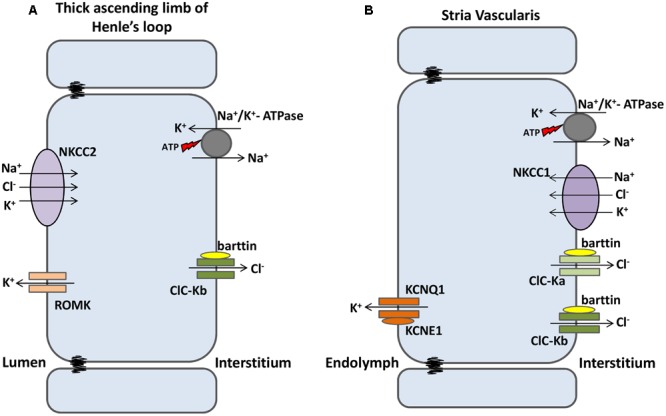
    1. Adachi S., Uchida S., Ito H., Hata M., Hiroe M., Marumo F., et al. (1994). Two isoforms of a chloride channel predominantly expressed in thick ascending limb of Henle’s loop and collecting ducts of rat kidney. J. Biol. Chem. 269 17677–17683. - PubMed
    1. Adrian R. H., Bryant S. H. (1974). On the repetitive discharge in myotonic muscle fibres. J. Physiol. 240 505–515. 10.1113/jphysiol.1974.sp010620 - DOI - PMC - PubMed
    1. Al-Awqati Q. (1995). Chloride channels of intracellular organelles. Curr. Opin. Cell Biol. 7 504–508. 10.1016/0955-0674(95)80006-9 - DOI - PubMed

LinkOut - more resources