

The Citrus Flavanone Naringenin Protects Myocardial Cells against Age-Associated Damage
- PMID: 28386313
- PMCID: PMC5366223
- DOI: 10.1155/2017/9536148
The Citrus Flavanone Naringenin Protects Myocardial Cells against Age-Associated Damage
Abstract
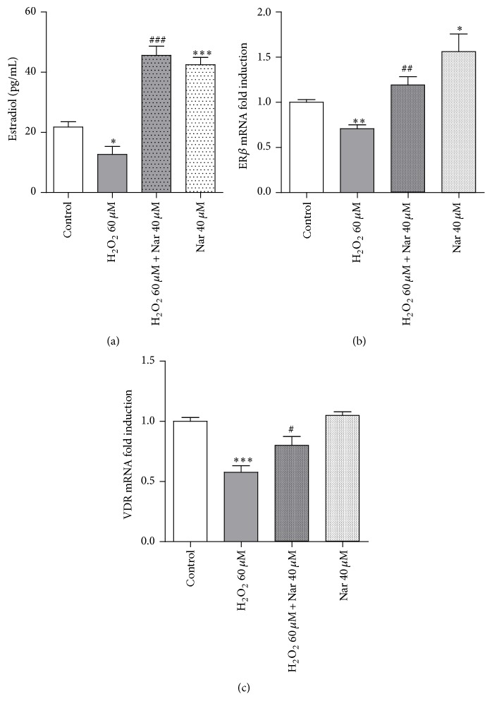
In recent years, the health-promoting effects of the citrus flavanone naringenin have been examined. The results have provided evidence for the modulation of some key mechanisms involved in cellular damage by this compound. In particular, naringenin has been revealed to have protective properties such as an antioxidant effect in cardiometabolic disorders. Very recently, beneficial effects of naringenin have been demonstrated in old rats. Because aging has been demonstrated to be directly related to the occurrence of cardiac disorders, in the present study, the ability of naringenin to prevent cardiac cell senescence was investigated. For this purpose, a cellular model of senescent myocardial cells was set up and evaluated using colorimetric, fluorimetric, and immunometric techniques. Relevant cellular senescence markers, such as X-gal staining, cell cycle regulator levels, and the percentage of cell cycle-arrested cells, were found to be reduced in the presence of naringenin. In addition, cardiac markers of aging-induced damage, including radical oxidative species levels, mitochondrial metabolic activity, mitochondrial calcium buffer capacity, and estrogenic signaling functions, were also modulated by the compound. These results suggested that naringenin has antiaging effects on myocardial cells.
Figures
Similar articles
-
Naringenin inhibits angiotensin II-induced vascular smooth muscle cells proliferation and migration and decreases neointimal hyperplasia in balloon injured rat carotid arteries through suppressing oxidative stress.Biol Pharm Bull. 2013;36(10):1549-55. doi: 10.1248/bpb.b13-00247. Epub 2013 Aug 3. Biol Pharm Bull. 2013. PMID: 23912743
-
Citrus flavanones naringenin and hesperetin improve antioxidant status and membrane lipid compositions in the liver of old-aged Wistar rats.Exp Gerontol. 2016 Nov;84:49-60. doi: 10.1016/j.exger.2016.08.014. Epub 2016 Aug 29. Exp Gerontol. 2016. PMID: 27587005
-
Effect of the citrus flavanone naringenin on oxidative stress in rats.J Agric Food Chem. 2007 Mar 21;55(6):2142-8. doi: 10.1021/jf061714h. Epub 2007 Feb 22. J Agric Food Chem. 2007. PMID: 17315884
-
Flavanones: Citrus phytochemical with health-promoting properties.Biofactors. 2017 Jul 8;43(4):495-506. doi: 10.1002/biof.1363. Epub 2017 May 12. Biofactors. 2017. PMID: 28497905 Review.
-
Combating oxidative stress disorders with citrus flavonoid: Naringenin.Life Sci. 2018 Sep 1;208:111-122. doi: 10.1016/j.lfs.2018.07.017. Epub 2018 Aug 10. Life Sci. 2018. PMID: 30021118 Review.
Cited by
-
Naringenin, a flavanone with antiviral and anti-inflammatory effects: A promising treatment strategy against COVID-19.Phytother Res. 2020 Dec;34(12):3137-3147. doi: 10.1002/ptr.6781. Epub 2020 Jul 2. Phytother Res. 2020. PMID: 32613637 Free PMC article. Review.
-
Naringenin Attenuates Myocardial Ischemia-Reperfusion Injury via cGMP-PKGIα Signaling and In Vivo and In Vitro Studies.Oxid Med Cell Longev. 2019 Jan 8;2019:7670854. doi: 10.1155/2019/7670854. eCollection 2019. Oxid Med Cell Longev. 2019. PMID: 30728891 Free PMC article.
-
Potential Role of Polyphenolic Flavonoids as Senotherapeutic Agents in Degenerative Diseases and Geroprotection.Pharmaceut Med. 2022 Dec;36(6):331-352. doi: 10.1007/s40290-022-00444-w. Epub 2022 Sep 13. Pharmaceut Med. 2022. PMID: 36100824 Free PMC article. Review.
-
Flavonoids-Natural Gifts to Promote Health and Longevity.Int J Mol Sci. 2022 Feb 16;23(4):2176. doi: 10.3390/ijms23042176. Int J Mol Sci. 2022. PMID: 35216290 Free PMC article. Review.
-
Nutritional components as mitigators of cellular senescence in organismal aging: a comprehensive review.Food Sci Biotechnol. 2022 Jun 18;31(9):1089-1109. doi: 10.1007/s10068-022-01114-y. eCollection 2022 Aug. Food Sci Biotechnol. 2022. PMID: 35756719 Free PMC article. Review.
References
MeSH terms
Substances
LinkOut - more resources
Full Text Sources
Other Literature Sources
