. 2017 Mar 15;9(3):1139-1150.
eCollection 2017.

Lipoxin A4 pretreatment mitigates skeletal muscle ischemia-reperfusion injury in rats

Affiliations

Lipoxin A4 pretreatment mitigates skeletal muscle ischemia-reperfusion injury in rats

Haiyang Zong et al. Am J Transl Res. .

Abstract

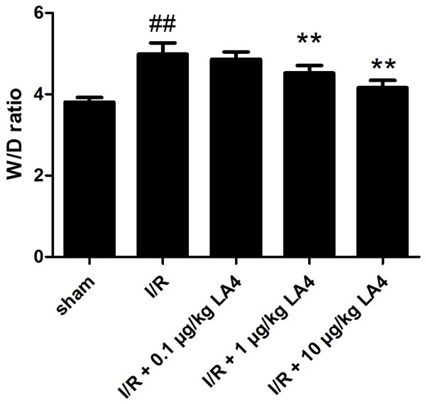
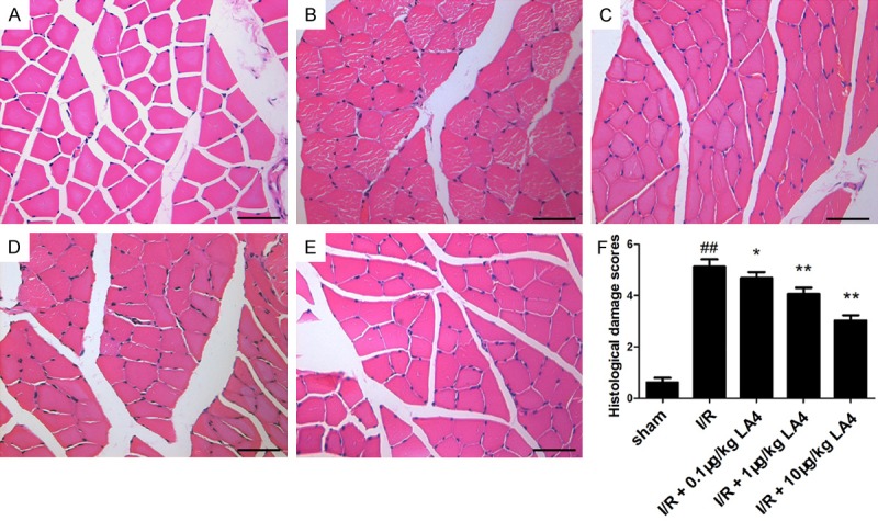
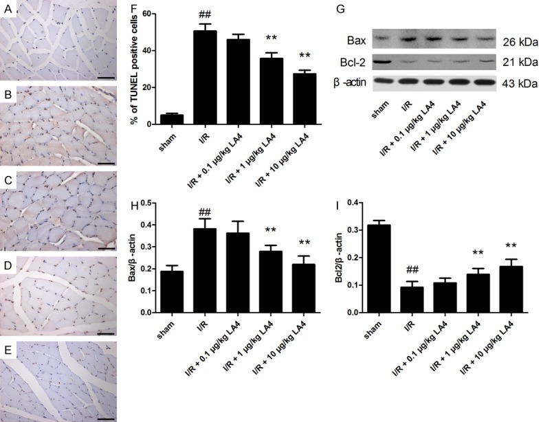
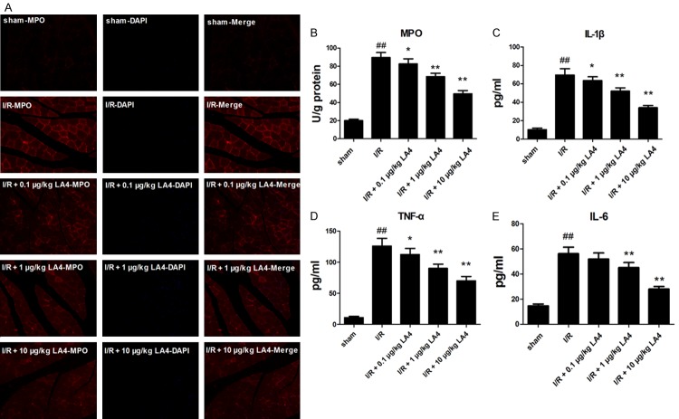
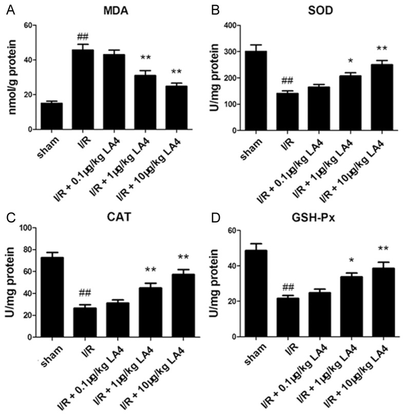
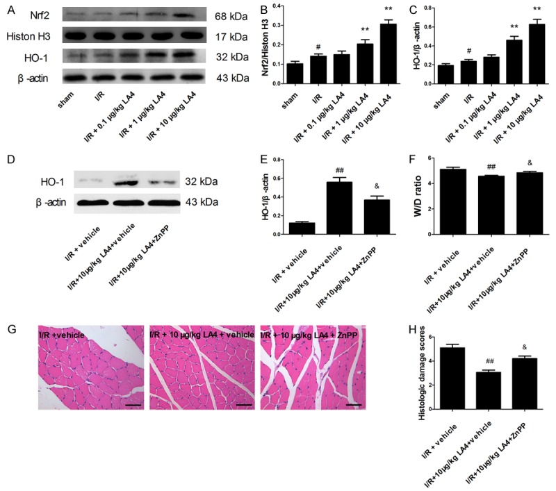
The aim of this study was to investigate the protective effects and underlying anti-oxidative molecular mechanism of lipoxin A4 (LA4) in rats with ischemia/reperfusion (I/R)-injured skeletal muscle. A rat model of I/R-injured skeletal muscle was obtained by subjecting rats to a 3-h ligation of the right femoral artery followed by 3 h of reperfusion. Treatment with LA4 significantly ameliorated histological damage scores in I/R-injured skeletal muscle. LA4 treatment resulted in remarkable decreases in the wet weight/dry weight ratio (W/D ratio), inflammatory response, oxidative stress, and cell apoptosis. In addition, treatment with LA4 was accompanied by a prominently enhanced nuclear accumulation of nuclear factor erythroid 2-related factor 2 (Nrf2) and expression of heme oxygenase 1 (HO-1) in the I/R-injured skeletal muscle. However, these protective effects were reversed by zinc protoporphyrin-IX (ZnPP), a specific HO-1 inhibitor. Our study shows that LA4 may have the potential as a therapeutic agent for I/R-injured muscle tissue via activation of the Nrf2/HO-1 signaling pathway.

Keywords: Ischemia-reperfusion injury; Nrf2/HO-1 signaling pathway; apoptosis; inflammatory response; lipoxin A4; oxidative stress; skeletal muscle.


Figures

Figure 1
Figure 1
Effects of administration of LA4 on tissue edema in skeletal muscle with I/R injury. The data are presented as mean ± SD. ##P < 0.01 compared with that of sham group; **P < 0.01 compared with that of I/R group.
Figure 2
Figure 2
Representative photomicrographs of skeletal muscle tissue following HE staining (scale bar = 50 μm). A. Sham group. B. I/R group. C. I/R + 0.1 μg/kg LA4 group. D. I/R + 1 μg/kg LA4 group. E. I/R + 10 μg/kg LA4 group. F. Quantification of histological damage scores in different groups. ##P < 0.01 compared with that of sham group; *P < 0.05 and **P < 0.01 compared with that of I/R group.
Figure 3
Figure 3
Protective effects of LA4 on muscle apoptosis in rats with I/R injury evaluated by TUNEL staining and Western blotting. (A) Sham group. (B) I/R group. (C) I/R + 0.1 μg/kg LA4 group. (D) I/R+ 1 μg/kg LA4 group. (E) I/R + 10 μg/kg LA4 group. (F) Quantification of apoptosis cells. (G) Blots of Bax and Bcl-2. (H, I) Semi-quantification of protein levels. Data are presented as mean ± SD. Scale bar = 50 μm (A-E). ##P < 0.01 compared with that of sham group; **P < 0.01 compared with that of I/R group.
Figure 4
Figure 4
Effect of LA4 treatment on I/R-injured skeletal muscle inflammatory responses. A. Immunofluorescent staining of MPO and DAPI in skeletal muscle sections (magnitude 200 ×) showing LA4 treatment decreased the number of MPO-positive cells. B-E. Changes in MPO activity and concentrations of TNF-α, IL-1β, and IL-6 in skeletal muscle tissue showing increased MPO activity, levels of TNF-α, IL-1β, and IL-6 in the muscle tissue after I/R injury, and significant reduction in this response following treatment with LA4. ##P < 0.01 compared with sham group; *P < 0.05 and **P < 0.01 compared with the I/R group.
Figure 5
Figure 5
Changes in MDA, SOD, CAT, and GSH-Px in muscle tissue (A-D). The content of MDA increases in the muscular tissue after I/R injury, the activities of SOD, CAT, and GSH-Px decrease, and LA4 treatment reverses these injury-induced changes. ##P < 0.01 compared with the sham group; *P < 0.05 and **P < 0.01 compared with the I/R group.
Figure 6
Figure 6
Involvement of activation of the Nrf2/HO-1 signaling pathway in the protective effects of LA4 in rats with skeletal muscle I/R injury. A. Protein expression of Nrf2 and HO-1. B, C. Quantification of Nrf2 and HO-1 levels showing LA4 dose dependently activated the Nrf2/HO-1 signaling pathway. D, E. ZnPP administered 1 h before ischemia and HO-1 protein expression determined by Western blotting. F. Skeletal muscle edema. G. Representative images of HE staining from I/R + vehicle group, I/R + 10 μg/kg LA4 + vehicle group, and I/R + 10 μg/kg LA4 + ZnPP group (scale bar = 50 μm). H. Histological damage scores. Results are expressed as mean ± SD. #P < 0.05 compared with sham group; **P < 0.01 compared with I/R group; ##P < 0.01 compared with I/R + vehicle group; &P < 0.05 compared with IR + 10 μg/kg LA4 + vehicle group.

References

    1. Wang WZ, Baynosa RC, Zamboni WA. Therapeutic interventions against reperfusion injury in skeletal muscle. J Surg Res. 2011;171:175–182. - PubMed
    1. Via AG, Oliva F, Spoliti M, Maffulli N. Acute compartment syndrome. Muscles Ligaments Tendons J. 2015;5:18–22. - PMC - PubMed
    1. Wang WZ, Baynosa RC, Zamboni WA. Update on ischemia-reperfusion injury for the plastic surgeon: 2011. Plast Reconstr Surg. 2011;128:685e–692e. - PubMed
    1. Blaisdell FW. The pathophysiology of skeletal muscle ischemia and the reperfusion syndrome: a review. Cardiovasc Surg. 2002;10:620–630. - PubMed
    1. Lindsay TF, Liauw S, Romaschin AD, Walker PM. The effect of ischemia reperfusion on adenine-nucleotide metabolism and xanthine-oxidase production in skeletal-muscle. J Vasc Surg. 1990;12:8–15. - PubMed

LinkOut - more resources