Review
. 2017 May;133(5):665-704.
doi: 10.1007/s00401-017-1707-9. Epub 2017 Apr 6.

Roles of tau protein in health and disease

Affiliations
Review

Roles of tau protein in health and disease

Tong Guo et al. Acta Neuropathol. 2017 May.

Abstract

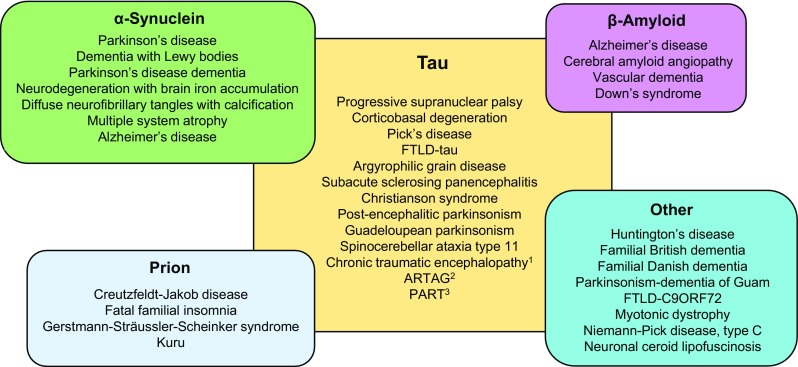
Tau is well established as a microtubule-associated protein in neurons. However, under pathological conditions, aberrant assembly of tau into insoluble aggregates is accompanied by synaptic dysfunction and neural cell death in a range of neurodegenerative disorders, collectively referred to as tauopathies. Recent advances in our understanding of the multiple functions and different locations of tau inside and outside neurons have revealed novel insights into its importance in a diverse range of molecular pathways including cell signalling, synaptic plasticity, and regulation of genomic stability. The present review describes the physiological and pathophysiological properties of tau and how these relate to its distribution and functions in neurons. We highlight the post-translational modifications of tau, which are pivotal in defining and modulating tau localisation and its roles in health and disease. We include discussion of other pathologically relevant changes in tau, including mutation and aggregation, and how these aspects impinge on the propensity of tau to propagate, and potentially drive neuronal loss, in diseased brain. Finally, we describe the cascade of pathological events that may be driven by tau dysfunction, including impaired axonal transport, alterations in synapse and mitochondrial function, activation of the unfolded protein response and defective protein degradation. It is important to fully understand the range of neuronal functions attributed to tau, since this will provide vital information on its involvement in the development and pathogenesis of disease. Such knowledge will enable determination of which critical molecular pathways should be targeted by potential therapeutic agents developed for the treatment of tauopathies.

Keywords: Alzheimer’s disease; Microtubule binding; Propagation; Synaptic dysfunction; Tau; Tauopathy.


Figures

Fig. 1
Fig. 1
Tau protein domains and alternative splicing in the human CNS. Six isoforms of tau are generated in the human CNS by alternative splicing of the MAPT gene. Distinct amino acid sequences encoded by exons 2 and 3 in the N-terminal region of tau are either excluded (0N), or differentially included giving rise to 1N (exon 2) or 2N (exons 2 and 3) tau isoforms. The central region of tau comprises the proline-rich domain (PRD). Alternative splicing of exon 10 in the microtubule binding domain (MTBD), results in 3R or 4R tau isoforms. The C-terminal region is common to all six human CNS tau isoforms. The actual molecular weight (MW, kDa), and the apparent (App.) MW of each tau isoform on SDS-PAGE, are indicated on the right
Fig. 2
Fig. 2
Binding of tau to microtubules. Tau associates with microtubules primarily through the microtubule binding domain, comprising either three or four repeats. The N and C termini of tau are closely associated when tau is free in the cytoplasm giving rise to the proposed “paper-clip” model of tau conformation. On binding to microtubules, the terminal regions of tau become separated and the N terminus of tau projects away from the microtubule surface
Fig. 3
Fig. 3
Post-translational modifications of tau. Illustration of the post-translational modifications identified on tau. The coloured bars indicate the approximate sites of each modification on the largest human CNS tau isoform (2N4R, 441 amino acids)
Fig. 4
Fig. 4
Tau localisation in neurons. Schematic depicts the differing locations of neuronal tau, the majority of which is associated with the microtubule cytoskeleton in axons. Tau is also located in the somatodendritic compartment, including in mitochondria, the nucleus, plasma membrane, and synapses. Dendritic tau (indicated in red) is increased in the tauopathies
Fig. 5
Fig. 5
Tauopathies. Diagram illustrating the wide range of neuropathological conditions in which tau pathology is a significant feature. The central panel illustrates disorders in which tau pathology is the primary feature. The overlapping panels summarise conditions in which tau inclusions are accompanied by deposits of other disease-associated proteins [19, 358, 445, 469]. 1Chronic traumatic encephalopathy includes traumatic brain injury and dementia pugilistica; 2ARTAG, aging-related tau astrogliopathy includes globular glial tauopathy; 3PART, primary age-related tauopathy includes tangle-predominant dementia and clinically asymptomatic cases; FTLD, frontotemporal lobar degeneration

References

    1. Aamodt EJ, Williams RC., Jr Biochemistry. 1984;23:6023–6031. doi: 10.1021/bi00320a019. - DOI - PubMed
    1. Abisambra JF, Jinwal UK, Blair LJ, O’Leary JC, 3rd, Li Q, Brady S, Wang L, Guidi CE, Zhang B, Nordhues BA, et al. J Neurosci. 2013;33:9498–9507. doi: 10.1523/JNEUROSCI.5397-12.2013. - DOI - PMC - PubMed
    1. Abraha A, Ghoshal N, Gamblin TC, Cryns V, Berry RW, Kuret J, Binder LI. J Cell Sci. 2000;113:3737–3745. - PubMed
    1. Ahmed T, Van der Jeugd A, Blum D, Galas MC, D’Hooge R, Buee L, Balschun D. Neurobiol Aging. 2014;35:2474–2478. doi: 10.1016/j.neurobiolaging.2014.05.005. - DOI - PubMed
    1. Akiyama H, Ikeda K, Kondo H, McGeer PL. Neurosci Lett. 1992;146:152–154. doi: 10.1016/0304-3940(92)90065-F. - DOI - PubMed

Publication types