. 2017 Dec;43(7):604-620.
doi: 10.1111/nan.12402. Epub 2017 May 15.

Novel conformation-selective alpha-synuclein antibodies raised against different in vitro fibril forms show distinct patterns of Lewy pathology in Parkinson's disease

Affiliations

Novel conformation-selective alpha-synuclein antibodies raised against different in vitro fibril forms show distinct patterns of Lewy pathology in Parkinson's disease

D J Covell et al. Neuropathol Appl Neurobiol. 2017 Dec.

Abstract

Aims: The aim of this study was to test the hypothesis that different conformations of misfolded α-synuclein (α-syn) are present in Parkinson's disease (PD) brain.

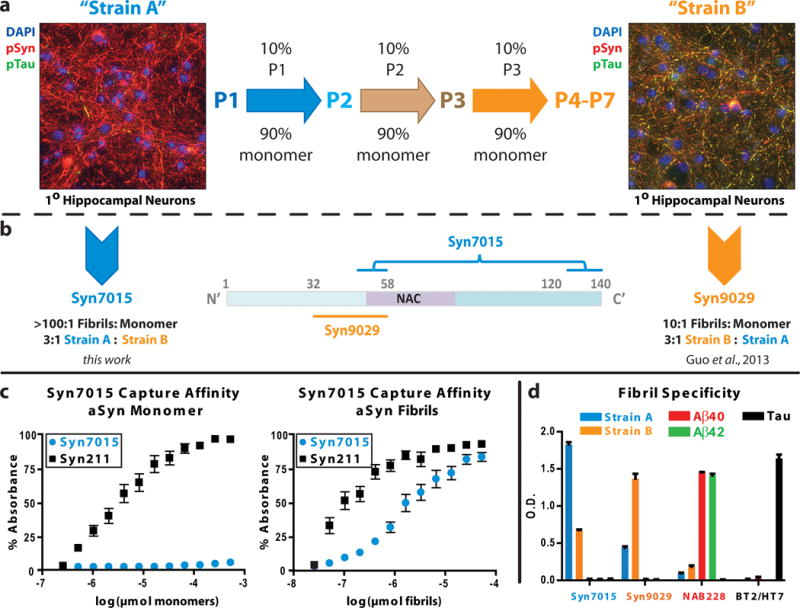
Methods: Using two previously characterized conformations of α-syn fibrils, we generated new conformation-selective α-syn monoclonal antibodies (mAbs). We then interrogated multiple brain regions in a well-characterized autopsy cohort of PD patients (n = 49) with these mAbs, Syn7015 and Syn9029.

Results: Syn7015 detects Lewy bodies (LBs) and Lewy neurites (LNs) formed by pathological α-syn in all brain regions tested, and is particularly sensitive to LNs and small Lewy dots, inclusions believed to form early in the disease. Further, we observed colocalization between Syn7015 and an early marker of α-syn pathology formation, phospho-Ser129-α-syn, and a lack of extensive colocalization with markers of more mature pathology. In comparison, Syn9029 detects Lewy pathology in all regions examined, but indicates significantly fewer LNs than Syn7015. In addition, colocalization of Syn9029 with later markers of α-syn pathology maturation (ubiquitin and P62) suggests that the pathology detected by Syn9029 is older. Semiquantitative scoring of both LN and LB pathology in nine brain regions further established this trend, with Syn7015 LN scores consistently higher than Syn9029 LN scores.

Conclusions: Our data indicate that different conformations of α-syn pathology are present in PD brain and correspond to different stages of maturity for Lewy pathology. Regional analysis of Syn7015 and Syn9029 immunostaining also provides support for the Braak hypothesis that α-syn pathology advances through the brain.

Keywords: Lewy pathology maturation; Parkinson's disease; amyloid; conformation-selective antibodies.


Conflict of interest statement

Conflict of Interest

All authors declare that they have no conflict of interest.

Figures

Figure 1
Figure 1
Generation of α-syn conforamtion selective mAbs. (a) De novo strain A (P1) PFFs induce extensive insoluble phospho-Ser129-α-syn (pSyn) positive aggregates (red) in wild-type mouse hippocampal neurons. Serially passaging (center) using 10% PFFs from the prior passage to seed monomeric α-syn generated strain B (P4-P7) α-syn fibrils, which induce both insoluble pSyn (red) and phosphorylated tau or pTau (green) in primary neuronal cultures (right). The aggregates are predominantly found in neuronal process where pSyn and pTau aggregates frequently co-localize (yellow). (b) Syn7015 recognizes a discontinuous epitope with binding elements in the extreme C-terminus and near amino acid 50 whereas Syn9029 recognizes a sequence between amino acid residues 32 and 58. The brackets connecting the two stretches of α-syn are intended to signify the discontinuous epitope recognized by Syn7015. (c) Syn7015 is highly selective for strain A fibrils over α-syn monomer when used as a capture antibody in sandwich ELISA, as compared to the control mAb, Syn211. (d) Comparison of the fibril binding affinity by direct ELISA of Syn7015 and Syn9029 for strain A and strain B α-syn fibrils, Aβ40 and Aβ42 fibrils, and tau fibrils. NAB228 and BT2/HT7 were used as Aβ and tau control mAbs.
Figure 2
Figure 2
Subcortical Lewy body (LB) and Lewy neurite (LN) pathology as detected by α-syn conformation-selective antibodies. Syn303, Syn7015 and Syn9029 each have a strong preference for pathological α-syn in (a–c) the dorsal motor nucleus of the medulla (d–f) the substantia nigra pars compacta, and (g–i) the central nucleus of the amygdala. Images are taken from serial sections. Bar = 100 μm. Representative LBs (black arrowheads), thick LNs (black double-arrows), thread LNs (red arrowheads), and dot LN pathology (red double-arrows) are shown.
Figure 3
Figure 3
Cortical Lewy body (LB) and Lewy neurite (LN) pathology as detected by α-syn conformation-selective antibodies. (a–c) Syn7015 reveals significantly more LNs than Syn303 or Syn9029 in the hippocampus CA2. (d–f) In the entorhinal cortex on the same section, LBs and LNs are readily detected by Syn303 and Syn7015, while Syn9029 stains LBs, but not LNs. (g–i) Anterior cingulate gyrus is frequently filled with LNs, including thread and dot LNs, as indicated by staining with Syn7015 in addition to the severe pathology already detected by Syn303. Syn9029 pathology is limited to a subset of the LBs and LNs. Images are taken from serial sections. Bar = 100 μm. Representative LBs (black arrowheads), thick LNs (black double-arrows), thread LNs (red arrowheads), and dot LN pathology (red double-arrows) are shown.
Figure 4
Figure 4
Specific post-translational markers of Lewy pathology maturity variably co-localize with α-syn conformation-selective antibodies. pSyn pathology co-localized with (a) Syn7015 positive thread and dot LNs and LBs and (b) Syn9029 LNs and LBs. Ubiquitination is limited to thick LNs and LBs and rarely co-localizes with (c) Syn7015 thread or dot LN pathology, while frequently co-localizing with (d) Syn9029 pathology. P62 is seen in a subset of α-syn LB pathology, a subset that is rarely (e) Syn7015 positive, but is (f) Syn9029 positive. Bar = 100 μm
Figure 5
Figure 5
Conformation-selective antibodies highlight α-syn pathology in early and late stages of Lewy pathology (LP) maturation. (a) Conformational mAbs Syn7015 and Syn9029 see pathological α-syn inclusions at different maturational stages in LP evolution or formation, based on immunofluoresence observations in PD brain. (b) Proposed LP maturation model, in which pSyn pathology is first readily detected by Syn7015 in longitudinally transversely sectioned axonal processes. Subsequently, a portion of these α-syn inclusions undergo a conformational compaction into denser structures including thick Lewy neurites and Lewy bodies, in neuronal perikarya that become detectable by Syn9029, as well as by antibodies to ubiquitin and P62.
Figure 6
Figure 6
Distribution plots of semi-quantitative scores demonstrate a preferential staining of Lewy neurites (LNs) by Syn7015 that is maintained throughout the disease course. Semi-quantitative Lewy body (LB) and LN scores for Syn303, Syn7015, and Syn9029 in the (a) substantia nigra and (b) amygdala show equivalent distribution for LBs but not LNs. Distribution plots of semi-quantitative LB and LN scores in the anterior cingulate gyrus for patients classified as (c) Braak PD stage 4, (d) Braak PD stage 5 (e) and Braak PD stage 6 suggest the relative patterns of LB and LN staining are preserved throughout disease progression.
Figure 7
Figure 7
Syn7015 detects additional pathological burden as compared to Syn9029 in Alzheimer’s disease (AD) with amygdala predominant Lewy pathology (LP). In AD cases with amygdala-predominant LP, α-syn Lewy body (LB) and Lewy neurite (LN) pathology is abundant and recognized by (a) Syn303 and (b) Syn7015, but (c) Syn9029 stains only LBs. Bar = 100 μm.

Comment in

References

    1. Forman MS, Trojanowski JQ, Lee VMY. Neurodegenerative diseases: a decade of discoveries paves the way for therapeutic breakthroughs. Nat Med. 2004;10:1055–63. - PubMed
    1. Braak H, Sandmann-Keil D, Gai W, Braak E. Extensive axonal Lewy neurites in Parkinson’s disease: a novel pathological feature revealed by alpha-synuclein immunocytochemistry. Neurosci Lett. 1999;265:67–9. - PubMed
    1. Spillantini MG, Schmidt ML, Lee VMY, Trojanowski JQ, Jakes R, Goedert M. alpha-Synuclein in Lewy bodies. Nature. 1997;388:839–40. - PubMed
    1. Breydo L, Wu JW, Uversky VN. α-Synuclein misfolding and Parkinson’s disease. Biochim Biophys Acta. 2012;1822:261–85. - PubMed
    1. Luna E, Luk KC. Bent out of shape: α-Synuclein misfolding and the convergence of pathogenic pathways in Parkinson’s disease. FEBS Lett. 2015;589:3749–59. - PMC - PubMed