. 2017 Apr 7:7:45740.
doi: 10.1038/srep45740.

Calcium oxalate crystals and oxalate induce an epithelial-to-mesenchymal transition in the proximal tubular epithelial cells: Contribution to oxalate kidney injury

Affiliations

Calcium oxalate crystals and oxalate induce an epithelial-to-mesenchymal transition in the proximal tubular epithelial cells: Contribution to oxalate kidney injury

Marcia Bastos Convento et al. Sci Rep. .

Abstract

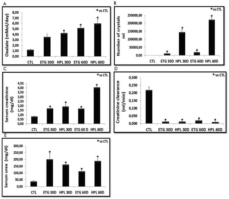
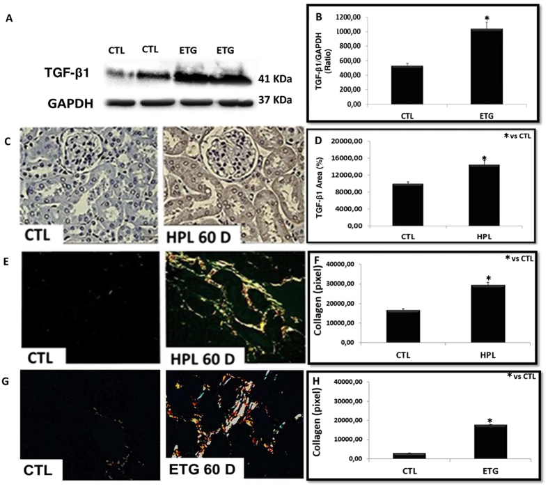
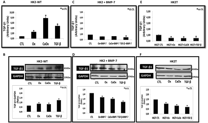
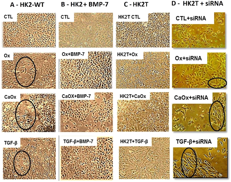
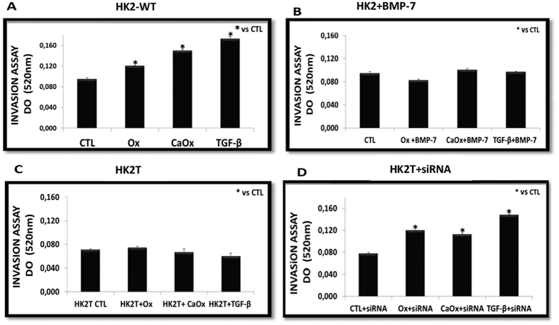
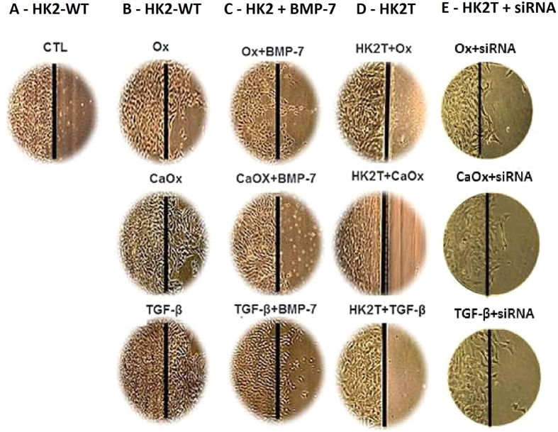
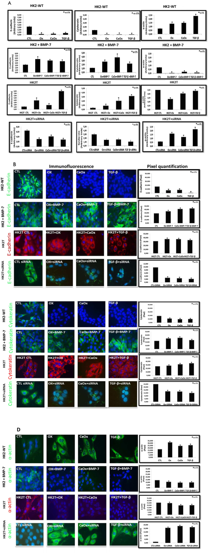
TGF-β1 is the main mediator of epithelial-to-mesenchymal transition (EMT). Hyperoxaluria induces crystalluria, interstitial fibrosis, and progressive renal failure. This study analyzed whether hyperoxaluria is associated with TGF-β1 production and kidney fibrosis in mice and if oxalate or calcium oxalate (CaOx) could induce EMT in proximal tubule cells (HK2) and therefore contribute to the fibrotic process. Hyperoxaluria was induced by adding hydroxyproline and ethylene glycol to the mice's drinking water for up to 60 days. Renal function and oxalate and urinary crystals were evaluated. Kidney collagen production and TGF-β1 expression were assessed. EMT was analyzed in vitro according to TGF-β1 production, phenotypic characterization, invasion, cell migration, gene and protein expression of epithelial and mesenchymal markers. Hyperoxaluric mice showed a decrease in renal function and an increase in CaOx crystals and Ox urinary excretion. The deposition of collagen in the renal interstitium was observed. HK2 cells stimulated with Ox and CaOx exhibited a decreased expression of epithelial as well as increased expression mesenchymal markers; these cells presented mesenchymal phenotypic changes, migration, invasiveness capability and TGF-β1 production, characterizing EMT. Treatment with BMP-7 or its overexpression in HK2 cells was effective at preventing it. This mechanism may contribute to the fibrosis observed in hyperoxaluria.


Conflict of interest statement

The authors declare no competing financial interests.

Figures

Figure 1
Figure 1
Animals were stimulated with hydroxyproline (HPL) and ethylene glycol (ETG) during 30 (30D) or 60 (60D) days increased oxalate excretion (A) and increased urinary crystals number (Neubauer camera count) (B). Impaired renal function has been demonstrated through increased serum creatinine (C), decreased of creatinine clearance (D), and increased serum urea (E). Control values represent the average of the data for the control group. Data are presented as means ± standard errors. N = 5 for each group. (*) Indicates significant differences compared with the control group at p < 0.05.
Figure 2
Figure 2. Hyperoxaluria increased the expression of renal fibrosis markers.
The stimulation with ethylene glycol during 60 days (ETG 60 D) and hydroxyproline during 60 days (HPL 60 D) increased the expression of TGF-β1 according to immunoblot image (A) and percentage of immunostaining area (C). There was an increase in the production of collagen type I (yellow to red tone) and collagen type III (greenish tone), showed by picrosirius red staining (E and G). Densitometric quantification of western blot bands (B), immunohistochemistry positive area (D) and collagen staining by picrosirius red (F and H) using ImageJ software. There was a significant increase in TGF-β1 protein expression and collagen production in HPL and ETG treated animals in comparison to controls (CTL) animals. Data are presented as means ± standard errors. N = 5 for each group. (*) Indicates significant differences compared with the control group at p < 0.05.
Figure 3
Figure 3
Wild type HK2 (HK2-WT) cells stimulated with potassium oxalate (Ox), calcium oxalate (CaOx), and TGF-β1 increased expression of TGF-β1 (A and B), while HK2 cells receiving BMP-7 (HK2 + BMP-7) (C and D) and HK2 overexpressing BMP-7 (HK2T) (E and F) did not increase TGF-β1 expression when compared to the control situation. Quantitative PCR analysis showing TGF-β1 mRNA expression in comparison to control situation (A,C and E). Western blot analysis and densitometric quantification using ImageJ software showing TGF-β1 protein expression in comparison to controls (CTL) (B,D and F). Data are presented as means ± standard errors. N = 15 for each group. (*) Indicates significant differences compared with the control group at p < 0.05.
Figure 4
Figure 4. Epithelial cell culture showing monolayer growth with clear and rounded delimitations.
On the contrary, mesenchymal cells lose cellular adhesion and present long, tuned and scattered morphology (black circle). Representative light microscopy images showing phenotypical changes in the HK2-WT (A), HK2 treated previously with BMP-7 (B), HK2 overexpressing BMP-7 (HK2T) (C) and HK2T and instable transfected with BMP-7 siRNA (HK2T + siRNA) (D). HK2 cells transfected or not were stimulated with potassium oxalate (Ox), calcium oxalate (CaOx) and TGF-β1 and compared to HK2 in control situation (CTL). On the contrary, HK2 + BMP-7 or HK2T showed only few or no morphological differentiations even in the presence of Ox, CaOx, and TGF-β1 in comparison to controls (CTL) situation. N = 15 for each group.
Figure 5
Figure 5. As a result of loss of cell adhesion and morphological changes from cuboid to fusiform form, these cells acquire the ability to invade (transwell chamber assays) and move in the extracellular matrix.
The HK2-WT (A) and HK2T + siRNA (D) acquired invasive capacity, while HK2 cells receiving BMP-7 (HK2 + BMP-7) (B), and HK2 cells overexpressing BMP-7 (HK2T) (C) behaved similarly the control (CTL) situation even in cell culture exposed to potassium oxalate (Ox), calcium oxalate (CaOx) and TGF-β1. Results were expressed as optical density (OD) and (*) groups were significantly different from the control situation (p < 0.05). N = 15 for each group.
Figure 6
Figure 6. Another characteristic of mesenchymal cell is the acquisition of migration ability.
Through light microscopy images we can observe that HK2-WT cells in situation control (A), even in confluence, do not migrate to the adjacent region of the culture plate. However stimulated with potassium oxalate (Ox), calcium oxalate (CaOx) and TGF-β1, HK2-WT cells and HK2T + siRNA migrate to the adjacent region (B and E). However, HK2 cells receiving BMP-7 (HK2 + BMP-7) (C) and HK2 cells overexpressing BMP-7 (HK2T) (D) behaved similarly to the control (CTL) situation. N = 15 for each group.
Figure 7
Figure 7. Demonstration of epithelial-to-mesenchymal transition event through epithelial (E-cadherin and Cytokeratin) and mesenchymal markers (smooth muscle α-actin).
Quantitative PCR analysis (A) and immunofluorescence images (FITC: green fluorescence, TRITC: red fluorescence and blue: nuclei) with their respective densitometric quantification using ImageJ software (B,C,D). We observed an increase in mesenchymal (smooth muscle α-actin) and a decrease in epithelial (cytokeratin and e-cadherin) marker expressions and fluorescence intensity in comparison to the control (CTL) situation in HK2-WT and HK2T + siRNA cells stimulated with potassium oxalate (Ox), calcium oxalate (CaOx) and TGF-β1. On the contrary, in HK2 receiving BMP-7 (HK2 + BMP-7) and HK2 cells overexpressing BMP-7 (HK2T) we did not observe a increase in mesenchymal (smooth muscle α-actin) and a decrease in epithelial (Cytokeratin and E-cadherin) markers. Data are presented as means ± standard errors. N = 15 for each group. (*) Significant different when compared to the control group at p < 0.05.

References

    1. Miller N. L., Evan A. P. & Lingeman J. E. Pathogenesis of Renal Calculi. Urologic Clinics of North America 34, 295–313 (2007). - PubMed
    1. Coe F. L., Evan A. & Worcester E. Kidney stone disease. J. Clin. Invest. 115, 2598–608 (2005). - PMC - PubMed
    1. Zeisberg M. & Kalluri R. The role of epithelial-to-mesenchymal transition in renal fibrosis. Journal of Molecular Medicine 82, 175–181 (2004). - PubMed
    1. Kalluri R. & Neilson E. G. Epithelial-mesenchymal transition and its implications for fibrosis. Journal of Clinical Investigation 112, 1776–1784 (2003). - PMC - PubMed
    1. Liu Y. Epithelial to mesenchymal transition in renal fibrogenesis: pathologic significance, molecular mechanism, and therapeutic intervention. J. Am. Soc. Nephrol. 15, 1–12 (2004). - PubMed

Publication types

MeSH terms

LinkOut - more resources