. 2017 Apr 7:7:46068.
doi: 10.1038/srep46068.

Targeting microbial biofilms using Ficin, a nonspecific plant protease

Affiliations

Targeting microbial biofilms using Ficin, a nonspecific plant protease

Diana R Baidamshina et al. Sci Rep. .

Abstract

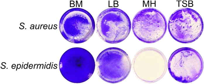
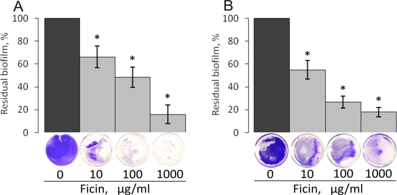
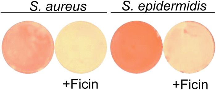
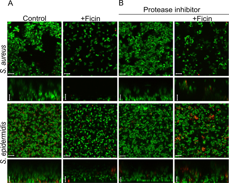
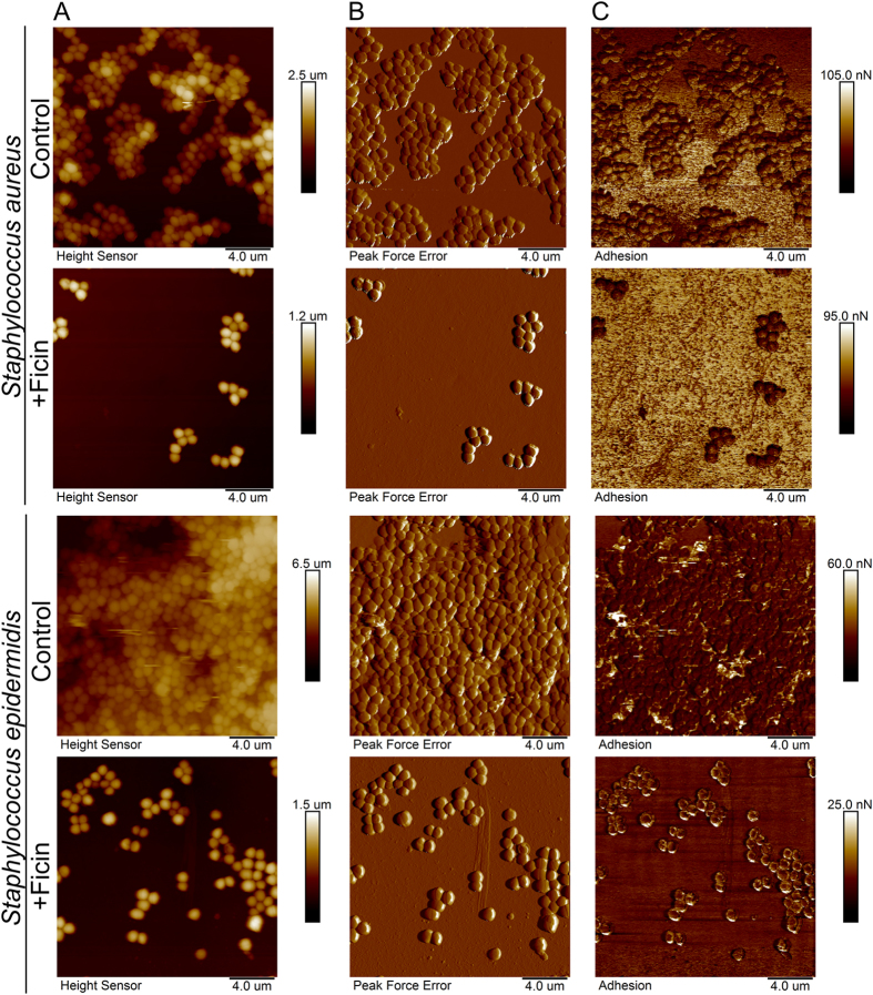
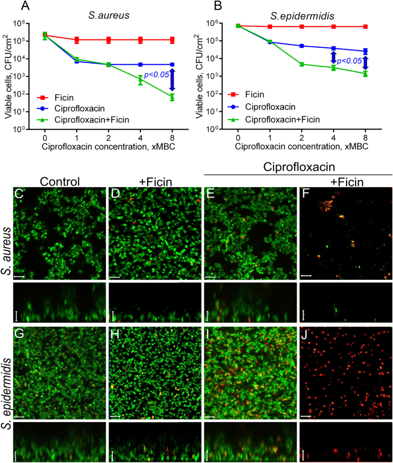
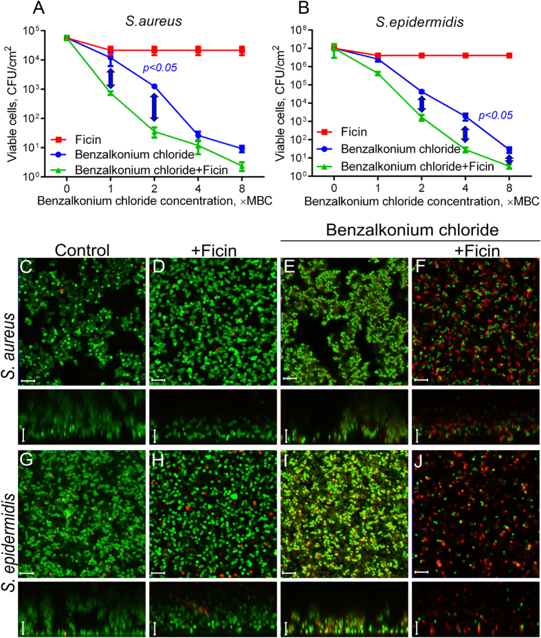
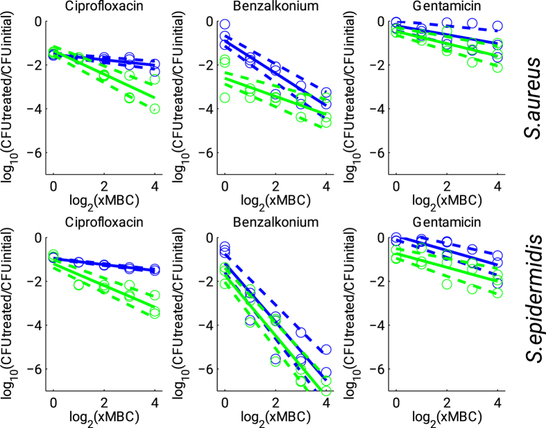
Biofilms, the communities of surface-attached bacteria embedded into extracellular matrix, are ubiquitous microbial consortia securing the effective resistance of constituent cells to environmental impacts and host immune responses. Biofilm-embedded bacteria are generally inaccessible for antimicrobials, therefore the disruption of biofilm matrix is the potent approach to eradicate microbial biofilms. We demonstrate here the destruction of Staphylococcus aureus and Staphylococcus epidermidis biofilms with Ficin, a nonspecific plant protease. The biofilm thickness decreased two-fold after 24 hours treatment with Ficin at 10 μg/ml and six-fold at 1000 μg/ml concentration. We confirmed the successful destruction of biofilm structures and the significant decrease of non-specific bacterial adhesion to the surfaces after Ficin treatment using confocal laser scanning and atomic force microscopy. Importantly, Ficin treatment enhanced the effects of antibiotics on biofilms-embedded cells via disruption of biofilm matrices. Pre-treatment with Ficin (1000 μg/ml) considerably reduced the concentrations of ciprofloxacin and bezalkonium chloride required to suppress the viable Staphylococci by 3 orders of magnitude. We also demonstrated that Ficin is not cytotoxic towards human breast adenocarcinoma cells (MCF7) and dog adipose derived stem cells. Overall, Ficin is a potent tool for staphylococcal biofilm treatment and fabrication of novel antimicrobial therapeutics for medical and veterinary applications.


Conflict of interest statement

The authors declare no competing financial interests.

Figures

Figure 1
Figure 1. The biofilm formation by S. aureus and S. epidermidis cultivated in Basal medium (BM), Luria-Bertani broth (LB), Müller-Hinton broth (MH), or Trypticase soy broth (TSB) on 35-mm polystyrol adhesive plates.
72 hours-old biofilms were stained by crystal violet.
Figure 2
Figure 2. The biofilm disruption by Ficin.
S. aureus (A) and S. epidermidis (B) were grown in BM broth for 72 h to form a rigid biofilm, the mature biofilms were gently washed by BM and a fresh BM broth was loaded. Ficin was added until final concentrations of 10, 100 or 1000 μg/ml and incubation was followed for 24 h. The residual biofilms were quantified by crystal-violet staining.
Figure 3
Figure 3. Evaluation of matrix proteins hydrolysis with Ficin.
Bacteria were grown on BM medium for 72 h to form a biofilm, then a medium was replaced by the fresh one containing Ficin (1000 μg/ml) and Congo red and incubation was continued for the next 24 h.
Figure 4
Figure 4. Confocal laser scanning microscopy.
S. aureus and S. epidermidis 48 h-old biofilms were established in cell imaging cover slips (Eppendorf) and treated with Ficin in absence (A) or presence (B) of protease inhibitors. After 24 h incubation cells were stained with DioC6 and propidium iodide to evaluate the cell viability. The scale bars indicate 5 µm.
Figure 5
Figure 5. Atomic force microscopy (Peak Force Tapping mode) of intact and Ficin-treated S. aureus and S. epidermidis biofilms.
Bacteria were grown in BM broth for 72 h to form a rigid biofilm, the mature biofilms were gently washed by BM and a fresh BM broth was loaded. Ficin was added until final concentrations of 1000 μg/ml and incubation was followed for 24 h. The residual biofilms were washed, fixed with glutardialdehyde and analyzed with AFM. (A) – height (topography); (B) – peak force error image; (C) –adhesion force image.
Figure 6
Figure 6. The Ficin treatment increases the efficacy of ciprofloxacin against biofilm-embedded Staphylococci.
Ficin (1000 μg/ml) and ciprofloxacin (1–8 × MBC) were added to 48 hours-old biofilms of S. aureus and S. epidermidis. After 24 h incubation, the biofilms were washed twice with sterile 0.9% NaCl. The adherent cells were scratched, resuspended and their viability was analyzed by using drop plate assay (A,B). Alternatively, 48 hours-old biofilms of S. aureus and S. epidermidis were incubated 24 h in presence of Ficin (1000 μg/ml) and ciprofloxacin (8 × MBC) in cell imaging coverglass slides and analyzed with confocal scanning microscopy (CJ). Significant differences between 10 log10 of the viable cell counts after treatment with ciprofloxacin in either absence of presence of Ficin according to Pearson’s Chi-squared homogeneity test (p < 0.05) are indicated in the figure. The scale bars indicate 5 µm.
Figure 7
Figure 7. The Ficin treatment increases the efficacy of benzalkonium chloride against biofilm-embedded Staphylococci.
Ficin (1000 μg/ml) and benzalkonium chloride (1–8 × MBC) were added to 48 hours-old biofilms of S. aureus and S. epidermidis. After 24 h incubation, the biofilms were washed twice with sterile 0.9% NaCl. The adherent cells were scratched, resuspended and their viability was analyzed by using drop plate assay (A,B). Alternatively, 48 hours-old biofilms of S. aureus and S. epidermidis were incubated 24 h in presence of Ficin (1000 μg/ml) and benzalkonium chloride (8 × MBC) in cell imaging coverglass slides and analyzed with confocal scanning microscopy (CJ). Significant differences between 10 log10 of the viable cell counts after treatment with benzalkonium chloride in either absence of presence of Ficin according to Pearson’s Chi-squared homogeneity test (p < 0.05) are indicated in the figure. The scale bars indicate 5 µm.
Figure 8
Figure 8. Dose-response curves for biofilm-embedded Staphylococci treated with antimicrobials in either presence (green) or absence (blue) of Ficin (1000 μg/ml).
Full lines denote regression lines, while dashed lines denote corresponding 95% confidence intervals.

References

    1. Yang L. et al.. Combating biofilms. Fems Immunology and Medical Microbiology 65, 146–157, doi: 10.1111/j.1574-695X.2011.00858.x (2012). - DOI - PubMed
    1. Richards J. J. & Melander C. Controlling Bacterial Biofilms. Chembiochem 10, 2287–2294, doi: 10.1002/cbic.200900317 (2009). - DOI - PubMed
    1. Donlan R. M. Biofilms: Microbial life on surfaces. Emerging Infectious Diseases 8, 881–890 (2002). - PMC - PubMed
    1. Worthington R. J., Blackledge M. S. & Melander C. Small-molecule inhibition of bacterial two-component systems to combat antibiotic resistance and virulence. Future Medicinal Chemistry 5, 1265–1284, doi: 10.4155/fmc.13.58 (2013). - DOI - PubMed
    1. Barth E., Myrvik Q. M., Wagner W. & Gristina A. G. In vitro and In vivo comparative colonization of staphylococcus-aureus and staphylococcus-epidermidis on orthopedic implant materials. Biomaterials 10, 325–328, doi: 10.1016/0142-9612(89)90073-2 (1989). - DOI - PubMed

Publication types

LinkOut - more resources