Review
. 2017 Jul;6(4):10.1002/wdev.272.
doi: 10.1002/wdev.272. Epub 2017 Apr 7.

Proximity-dependent labeling methods for proteomic profiling in living cells

Affiliations
Review

Proximity-dependent labeling methods for proteomic profiling in living cells

Chiao-Lin Chen et al. Wiley Interdiscip Rev Dev Biol. 2017 Jul.

Abstract

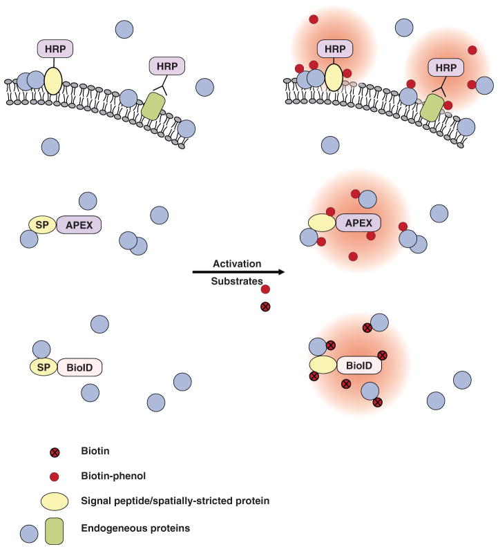
Characterizing the proteome composition of organelles and subcellular regions of living cells can facilitate the understanding of cellular organization as well as protein interactome networks. Proximity labeling-based methods coupled with mass spectrometry (MS) offer a high-throughput approach for systematic analysis of spatially restricted proteomes. Proximity labeling utilizes enzymes that generate reactive radicals to covalently tag neighboring proteins with biotin. The biotinylated endogenous proteins can then be isolated for further analysis by MS. To analyze protein-protein interactions or identify components that localize to discrete subcellular compartments, spatial expression is achieved by fusing the enzyme to specific proteins or signal peptides that target to particular subcellular regions. Although these technologies have only been introduced recently, they have already provided deep insights into a wide range of biological processes. Here, we describe and compare current methods of proximity labeling as well as their applications. As each method has its own unique features, the goal of this review is to describe how different proximity labeling methods can be used to answer different biological questions. WIREs Dev Biol 2017, 6:e272. doi: 10.1002/wdev.272 For further resources related to this article, please visit the WIREs website.


Conflict of interest statement

Conflict of interest: The authors have declared no conflicts of interest for this article.

Figures

FIGURE 1
FIGURE 1
Proximity labeling for proteomic profiling. To achieve regional protein labeling, the enzymes are usually fused with a targeting signal peptide or a spatially restricted protein (SP). The enzymes can also be fused with any protein of interest for protein interactome studies. After performing proximity labeling in living cells, the cells are lysed and the biotinylated endogenous proteins are isolated using steptavidin beads. Small peptides of enriched proteins are generated by trypsin digestion and subsequently ionized for mass spectrometry analysis. The mass-to-charge (m/z) ratio of each peptide is then used to identify peptide sequence usually through computational comparison against established databases.
FIGURE 2
FIGURE 2
Proximity labeling methods. BioID, a mutant form of the biotin ligase BirA, can convert biotin into radicals that can covalently tag neighboring proteins on lysine residues. HRP and APEX are peroxidases that, when activated by H2O2, are able to turn biotin-phenol substrates into highly reactive radicals that covalently tag neighboring proteins on electron-rich amino acids. In addition, fluorescein-aryl azide or biotin-aryl azide have been used for HRP-mediated proximity labeling (not shown in the figure). HRP is inactive in a reducing environment, such as the cytosol, but functions extracellularly. APEX, engineered ascorbate peroxidase; BioID, proximity-dependent biotin identification; HRP, horseradish peroxidase; SP, spatially restricted protein.

References

    1. Carrasco S, Meyer T. STIM proteins and the endoplasmic reticulum-plasma membrane junctions. Annu Rev Biochem. 2011;80:973–1000. doi: 10.1146/annurev-biochem-061609-165311. - DOI - PMC - PubMed
    1. Hogan PG, Lewis RS, Rao A. Molecular basis of calcium signaling in lymphocytes: STIM and ORAI. Annu Rev Immunol. 2010;28:491–533. doi: 10.1146/annurev.immunol.021908.132550. - DOI - PMC - PubMed
    1. Stefan CJ, Manford AG, Emr SD. ER-PM connections: sites of information transfer and inter-organelle communication. Curr Opin Cell Biol. 2013;25:434–442. doi: 10.1016/j.ceb.2013.02.020. - DOI - PMC - PubMed
    1. Elbaz Y, Schuldiner M. Staying in touch: the molecular era of organelle contact sites. Trends Biochem Sci. 2011;36:616–623. doi: 10.1016/j.tibs.2011.08.004. - DOI - PubMed
    1. Ong SE, Blagoev B, Kratchmarova I, Kristensen DB, Steen H, Pandey A, Mann M. Stable isotope labeling by amino acids in cell culture, SILAC, as a simple and accurate approach to expression proteomics. Mol Cell Proteomics. 2002;1:376–386. - PubMed

Publication types