

Intramuscular hemangioma causing periosteal reaction and cortical hypertrophy misdiagnosed as osteoid osteoma
- PMID: 28388514
- PMCID: PMC5384294
- DOI: 10.1016/j.ijscr.2017.03.013
Intramuscular hemangioma causing periosteal reaction and cortical hypertrophy misdiagnosed as osteoid osteoma
Abstract
Introduction: Intramuscular hemangioma in the periosteal region is rare. Although comprising less than 1% of all hemangiomas, they represent the most common type of intramuscular tumors. When located adjacent to bone, a periosteal reaction can occur. The deep localization of the hemangioma poses the diagnosis difficult. Only 8% to 19% of cases were diagnosed before surgery according to the literature review.
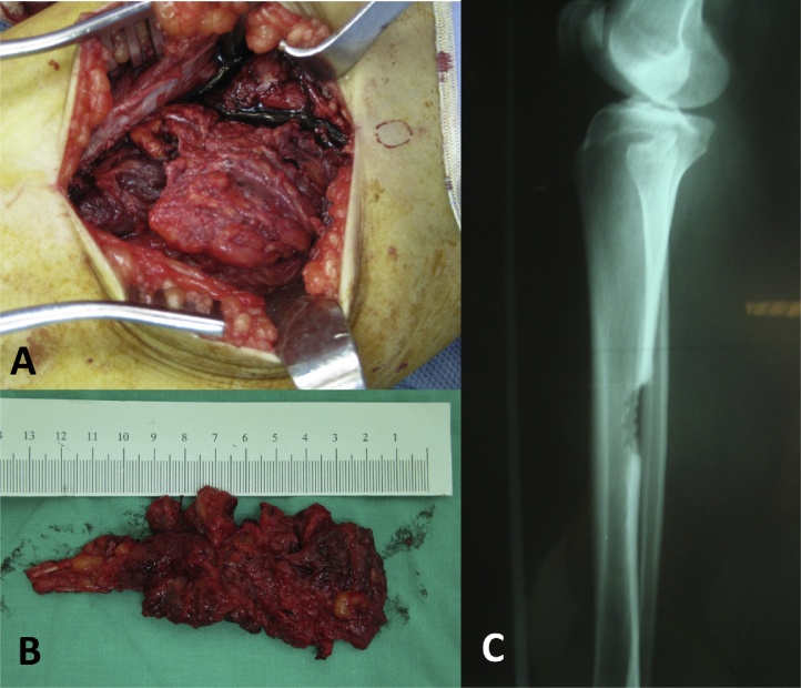
Presentation of case: We present a case of forty-one-year-old female diagnosed with intramuscular hemangioma, mimicking osteoid osteoma, adjacent to the periosteal region of tibia diaphysis treated by surgical excision.
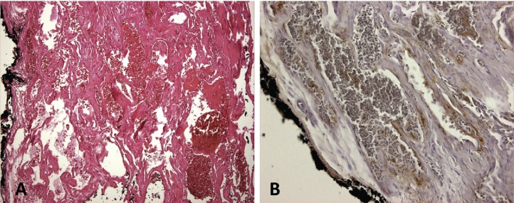
Discussion: When intramuscular hemangioma occurs nearby a bone structure, it can cause cortical, medullary and periosteal bone changes that are frequently misdiagnosed by plain radiography. Due to their infrequency, deep location, and atypical presentation, these lesions are seldom diagnosed at presentation. The hemangioma of the periosteal region can be locally destructive due to compression exerted on neighboring structures. It does not regress spontaneously, and surgical excision is frequently needed.
Conclusion: Intramuscular hemangioma of periosteal region occurs most commonly adjacent to long bones of the lower limb. They can cause hypertrophic periosteal reactions mimicking a periosteal or parosteal tumor. Although osteoid osteoma was considered in the differential diagnosis, MRI with enhancement should be performed to exclude intramuscular hemangioma. This may avoid unnecessary aggressive en-bloc tumor excisions resulting in bone weakness and prolonged rehabilitation. This case report has been written in line with the SCARE criteria (Agha et al., 2016 [1]).
Keywords: Intramuscular hemangioma; Osteoid osteoma; Periosteal reaction.
Copyright © 2017 The Authors. Published by Elsevier Ltd.. All rights reserved.
Figures


References
-
- Agha R.A. The SCARE statement: consensus-based surgical case report guidelines. Int. J. Surg. 2016;34:180–186. - PubMed
-
- Memis A. Magnetic resonance imaging of intramuscular haemangiomas with emphasis on contrast enhancement patterns. Clin. Radiol. 1996;51(3):198–204. - PubMed
-
- Welsh D., Hengerer A.S. The diagnosis and treatment of intramuscular hemangiomas of the masseter muscle. Am. J. Otolaryngol. 1980;1(2):186–190. - PubMed
-
- Goto T. Soft-tissue haemangioma and periosteal new bone formation on the neighbouring bone. Arch. Orthop. Trauma Surg. 2001;121(10):549–553. - PubMed
LinkOut - more resources
Full Text Sources
Other Literature Sources
Research Materials
