. 2017 May 23;8(21):34191-34204.
doi: 10.18632/oncotarget.16573.

Involvement of heparanase in the pathogenesis of acute kidney injury: nephroprotective effect of PG545

Affiliations

Involvement of heparanase in the pathogenesis of acute kidney injury: nephroprotective effect of PG545

Zaid Abassi et al. Oncotarget. .

Abstract

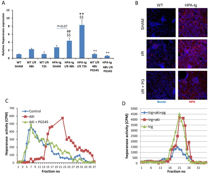
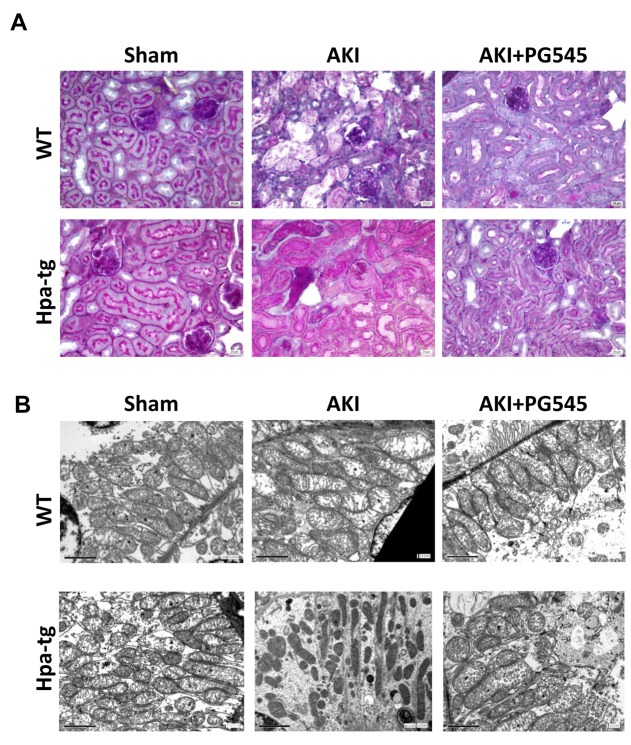
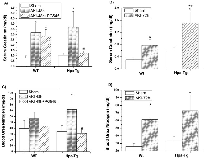
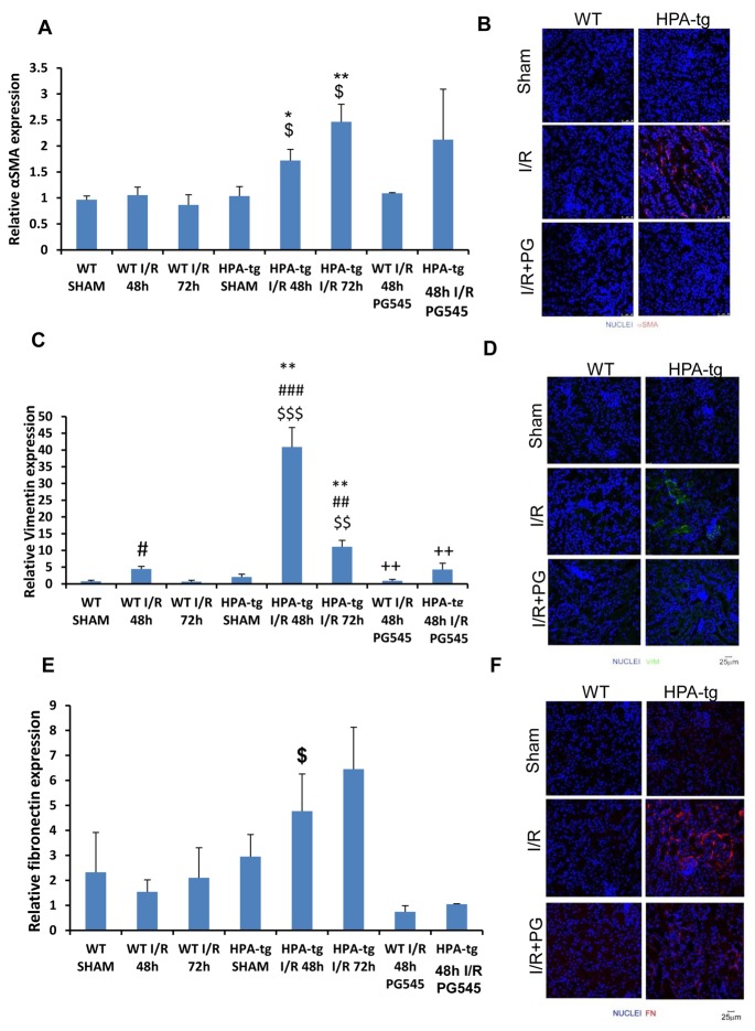
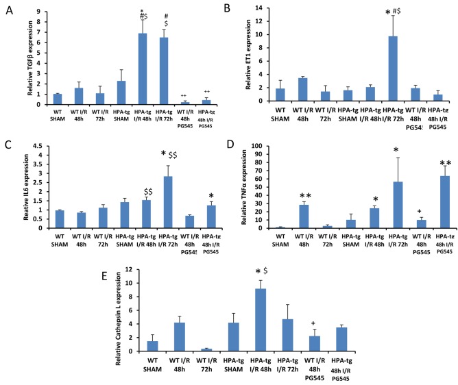
Despite the high prevalence of acute kidney injury (AKI) and its association with increased morbidity and mortality, therapeutic approaches for AKI are disappointing. This is largely attributed to poor understanding of the pathogenesis of AKI. Heparanase, an endoglycosidase that cleaves heparan sulfate, is involved in extracellular matrix turnover, inflammation, kidney dysfunction, diabetes, fibrosis, angiogenesis and cancer progression. The current study examined the involvement of heparanase in the pathogenesis of ischemic reperfusion (I/R) AKI in a mouse model and the protective effect of PG545, a potent heparanase inhibitor. I/R induced tubular damage and elevation in serum creatinine and blood urea nitrogen to a higher extent in heparanase over-expressing transgenic mice vs. wild type mice. Moreover, TGF-β, vimentin, fibronectin and α-smooth muscle actin, biomarkers of fibrosis, and TNFα, IL6 and endothelin-1, biomarkers of inflammation, were upregulated in I/R induced AKI, primarily in heparanase transgenic mice, suggesting an adverse role of heparanase in the pathogenesis of AKI. Remarkably, pretreatment of mice with PG545 abolished kidney dysfunction and the up-regulation of heparanase, pro-inflammatory (i.e., IL-6) and pro-fibrotic (i.e., TGF-β) genes induced by I/R. The present study provides new insights into the involvement of heparanase in the pathogenesis of ischemic AKI.Our results demonstrate that heparanase plays a deleterious role in the development of renal injury and kidney dysfunction,attesting heparanase inhibition as a promising therapeutic approach for AKI.

Keywords: PG545; acute kidney injury; heparanase; inflammation; ischemia.


Conflict of interest statement

CONFLICTS OF INTEREST

Edward Hammond is employed by Zucero Therapeutics, Darra, Queensland, Australia.

Figures

Figure 1
Figure 1. Heparanase regulation by I/R kidney injury
A. Bar plot showing relative gene expression of HPSE evaluated by real-time PCR in renal tissue extract from wt and Hpa-tg mice untreated or pre-treated with PG545. Results were normalized to GAPDH expression. B. Representative immunofluorescence staining of heparanase in cortical renal tissues of wt and Hpa-tg mice 48 h after I/R kidney injury, with or without pre-treatment with PG545. Magnification 40X. Representative heparanase activity in the renal tissue of wt C. and Hpa-tg D. mice prior and post I/R in the presence or absence of PG545 pretreatment. I/R, ischemia/reperfusion. *p < 0.05, **p < 0.01 vs. corresponding sham; #p < 0.05, ##p < 0.01 vs. corresponding group w/o I/R ; $p < 0.05, $$p < 0.01 vs. corresponding wt; +p < 0.05, ++p < 0.01 vs. untreated I/R corresponding group.
Figure 2
Figure 2. Ischemia/reperfusion (I/R) kidney injury in wt and Hpa-tg mice
I/R kidney injury was induced in wt and Hpa-tg mice by 30 minutes of clamping of both renal arteries. Mice were sacrificed after 48 hours. A. Representative images of PAS staining of paraffin-embedded cortex sections from various experimental groups. Magnification 40x. B. Electron microscopy micrographs of cortical renal tissue from wt and Hpa-tg mice that were subjected to 30 minutes of clamping of both renal arteries in the absence or presence of PG545 (0.4 mg/mouse, i.v). Note mitochondrial swelling and damage to mitochondrial cristae. Note normal ultrastructural appearance of mitochondria from mice treated with PG545. Magnification 12,000×.
Figure 3
Figure 3. Biomarkers of kidney function in wt and Hpa-Tg mice after I/R
Effect of AKI on serum creatinine (SCr) A., B. and Blood urea nitrogen (BUN) C., D. 48 h A., C. and 72 h B., D. after I/R insult in wt and Hpa-tg mice. Note that AKI induced a more profound increase in both SCr and BUN in Hpa-tg mice as compared with wt mice. Additional two groups of wt and Hpa-tg mice were pretreated one day prior to AKI induction with PG545 (0.4 mg/mouse, ip) and sacrificed 48 h after renal injury A., C. *p < 0.05, **p < 0.01 vs. sham; #p < 0.01 vs. untreated mice; $ p < 0.05 vs. wt.
Figure 4
Figure 4. Expression of epithelial mesenchymal transition (EMT) markers in wt vs
Hpa-tg mice after I/R kidney injury. Relative gene expression of A. α-SMA, C. VIM, and E. FN, were evaluated by real-time PCR in renal tissue extracts from wt and Hpa-tg mice that underwent I/R kidney injury with or without pre-treatment with PG545. Results were normalized to GAPDH expression. **p < 0.001 vs. wt SHAM mice. I/R, ischemia/reperfusion. Representative immunofluorescence staining of α-SMA B., VIM D. and FN F. in renal tissue of wt and Hpa-tg mice that underwent 48 h of I/R kidney injury with or without pre-treatment with PG545. Magnification 40x. *p < 0.05, **p < 0.01 vs. corresponding sham; #p < 0.05, ##p < 0.01 vs. corresponding group w/o I/R; $p < 0.05, $$p < 0.01 vs. corresponding wt; +p < 0.05, ++p < 0.01 vs. untreated I/R corresponding group.
Figure 5
Figure 5. TGF-β, ET-1, IL6, TNFα and cathepsin L gene expression in wt vs
Hpa-tg mice after I/R kidney injury. Bar plots representing relative gene expression of TGF-β A., ET-1B., IL6 C., TNFα D. and Cathepsin L E., evaluated by real-time PCR in renal tissue extracts from wt and Hpa-tg mice that underwent I/R kidney injury with or without pre-treatment with PG545. Results were normalized to GAPDH expression. *p < 0.05, **p < 0.001 vs. wt sham; #p < 0.05, ##p < 0.01 vs. corresponding group w/o I/R ; $p < 0.05, $$p < 0.01 vs. corresponding wt; +p < 0.05, ++p < 0.01 vs. untreated I/R corresponding group.

References

    1. Bonventre JV, Yang L. Cellular pathophysiology of ischemic acute kidney injury. J Clin Invest. 2011;121:4210–4221. - PMC - PubMed
    1. Schrier RW, Wang W, Poole B, Mitra A. Acute renal failure: definitions, diagnosis, pathogenesis, and therapy. J Clin Invest. 2004;114:5–14. - PMC - PubMed
    1. Lameire N, Van Biesen W, Vanholder R. Acute renal failure. Lancet. 2005;365:417–430. - PubMed
    1. Sheridan AM, Bonventre JV. Pathophysiology of ischemic acute renal failure. Contrib Nephrol. 2001;132:7–21. - PubMed
    1. Agarwal A, Nick HS. Renal response to tissue injury: lessons from heme oxygenase-1 GeneAblation and expression. J Am Soc Nephrol. 2000;11:965–973. - PubMed

MeSH terms

LinkOut - more resources