

FZUImageReg: A toolbox for medical image registration and dose fusion in cervical cancer radiotherapy
- PMID: 28388623
- PMCID: PMC5384778
- DOI: 10.1371/journal.pone.0174926
FZUImageReg: A toolbox for medical image registration and dose fusion in cervical cancer radiotherapy
Abstract
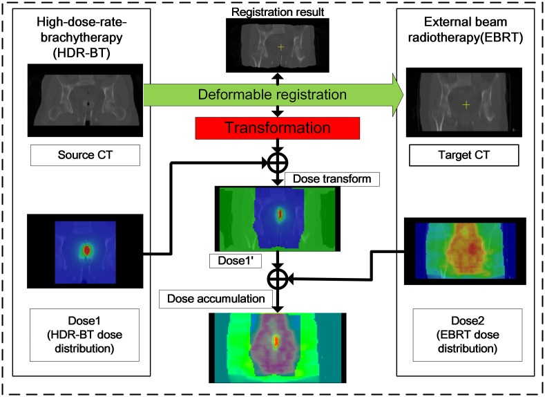
The combination external-beam radiotherapy and high-dose-rate brachytherapy is a standard form of treatment for patients with locally advanced uterine cervical cancer. Personalized radiotherapy in cervical cancer requires efficient and accurate dose planning and assessment across these types of treatment. To achieve radiation dose assessment, accurate mapping of the dose distribution from HDR-BT onto EBRT is extremely important. However, few systems can achieve robust dose fusion and determine the accumulated dose distribution during the entire course of treatment. We have therefore developed a toolbox (FZUImageReg), which is a user-friendly dose fusion system based on hybrid image registration for radiation dose assessment in cervical cancer radiotherapy. The main part of the software consists of a collection of medical image registration algorithms and a modular design with a user-friendly interface, which allows users to quickly configure, test, monitor, and compare different registration methods for a specific application. Owing to the large deformation, the direct application of conventional state-of-the-art image registration methods is not sufficient for the accurate alignment of EBRT and HDR-BT images. To solve this problem, a multi-phase non-rigid registration method using local landmark-based free-form deformation is proposed for locally large deformation between EBRT and HDR-BT images, followed by intensity-based free-form deformation. With the transformation, the software also provides a dose mapping function according to the deformation field. The total dose distribution during the entire course of treatment can then be presented. Experimental results clearly show that the proposed system can achieve accurate registration between EBRT and HDR-BT images and provide radiation dose warping and fusion results for dose assessment in cervical cancer radiotherapy in terms of high accuracy and efficiency.
Conflict of interest statement
Figures


Similar articles
-
Deformable image registration for dose mapping between external beam radiotherapy and brachytherapy images of cervical cancer.Phys Med Biol. 2019 May 31;64(11):115023. doi: 10.1088/1361-6560/ab1378. Phys Med Biol. 2019. PMID: 30913542
-
Evaluation of deformable image registration between external beam radiotherapy and HDR brachytherapy for cervical cancer with a 3D-printed deformable pelvis phantom.Med Phys. 2017 Apr;44(4):1445-1455. doi: 10.1002/mp.12168. Med Phys. 2017. PMID: 28214368
-
Anatomic structure-based deformable image registration of brachytherapy implants in the treatment of locally advanced cervix cancer.Brachytherapy. 2016 Sep-Oct;15(5):584-92. doi: 10.1016/j.brachy.2016.04.390. Epub 2016 Jun 1. Brachytherapy. 2016. PMID: 27263057
-
Dose Summation Strategies for External Beam Radiation Therapy and Brachytherapy in Gynecologic Malignancy: A Review from the NRG Oncology and NCTN Medical Physics Subcommittees.Int J Radiat Oncol Biol Phys. 2021 Nov 15;111(4):999-1010. doi: 10.1016/j.ijrobp.2021.06.019. Epub 2021 Jun 17. Int J Radiat Oncol Biol Phys. 2021. PMID: 34147581 Free PMC article. Review.
-
A review of segmentation and deformable registration methods applied to adaptive cervical cancer radiation therapy treatment planning.Artif Intell Med. 2015 Jun;64(2):75-87. doi: 10.1016/j.artmed.2015.04.006. Epub 2015 May 16. Artif Intell Med. 2015. PMID: 26025124 Review.
References
-
- Kamangar F, Dores GM, Anderson WF. Patterns of cancer incidence, mortality, and prevalence across five continents: defining priorities to reduce cancer disparities in different geographic regions of the world. Journal of clinical oncology. 2006;24(14):2137–50. 10.1200/JCO.2005.05.2308 - DOI - PubMed
-
- Siegel RL, Miller KD, Jemal A. Cancer statistics, 2016. CA: a cancer journal for clinicians. 2016;66(1):7–30. - PubMed
-
- Han K, Milosevic M, Fyles A, Pintilie M, Viswanathan AN. Trends in the utilization of brachytherapy in cervical cancer in the United States. International Journal of Radiation Oncology* Biology* Physics. 2013;87(1):111–9. - PubMed
-
- Yan D, Vicini F, Wong J, Martinez A. Adaptive radiation therapy. Physics in medicine and biology. 1997;42(1):123 - PubMed
MeSH terms
LinkOut - more resources
Full Text Sources
Other Literature Sources
Medical
